In a latest examine printed within the Journal of Biomedical Optics, researchers display the multimodal performance of focused ocular fluorescence spectroscopy in vitro and in vivo.
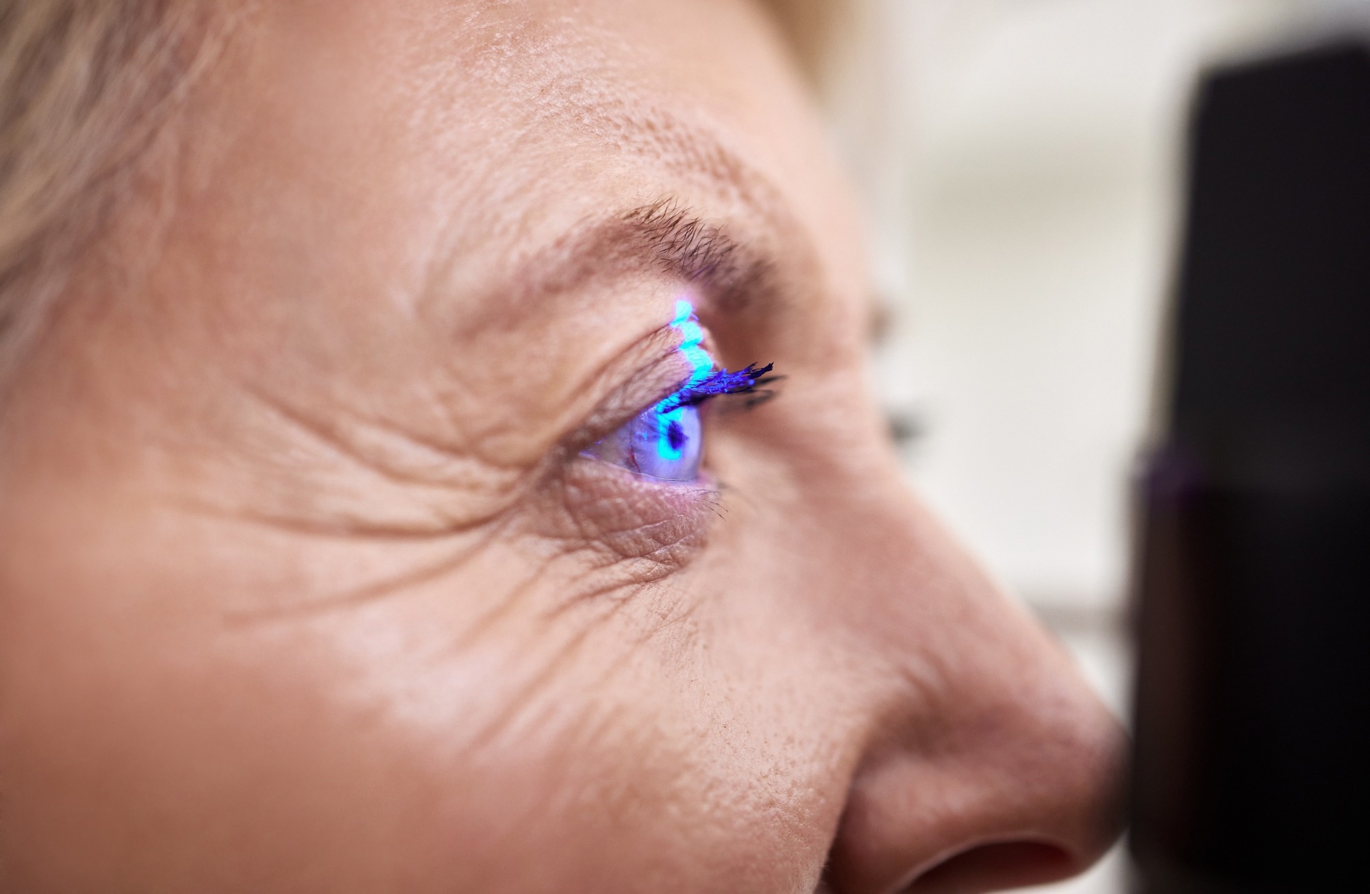 Examine: Focused spectroscopy within the eye fundus. Picture Credit score: PeopleImages.com – Yuri A / Shutterstock.com
Background
Some typical structural and practical modifications happen within the eyes, particularly the attention fundus, on account of ocular ailments like diabetic retinopathy (DR), age-related macular degeneration (AMD), and glaucoma. Neurological ailments like Alzheimer’s illness (AD) and Parkinson’s illness (PD) may also result in retinal modifications, resembling thinning of the retinal nerve fiber layer (RNFL) and modifications in hemodynamics.
Given the extremely heterogeneous options and composition of the attention fundus, biomarkers both get broadly dispersed all through this tissue or localize to particular areas. For instance, β-amyloid plaques unfold all through the retina of AD sufferers, whereas sufferers with DR have localized hemorrhages.
Typical imaging strategies don’t present sufficient information on retinal modifications induced by these ailments as in comparison with focused ocular diffuse reflectance spectroscopy (DRS). Ocular DRS strategies allow spectral evaluation of particular elements of the attention fundus, together with the optic disc, peripheral retina, and fovea between 500 and 800 nanometers (nm).
Diffuse reflectance and fluorescence spectroscopy may also elucidate the affect of things like lipofuscin accumulation, RNFL structural modifications, blood absorption spectrum, and melanin spectral profile, all of which affect the optical properties of retinal tissues.
In regards to the examine
Within the present examine, researchers determine the important thing options of the focused ocular spectroscopy know-how in vitro utilizing a reference goal and mannequin eye. The reference goal was an ultrahigh-definition display screen with a grid of eight totally different colours, with the fundus digicam positioned in entrance of it and solely amassing mild emitted by the display screen. The OEMI-7 eye mannequin, a seven mm pupil precisely simulating the human eye, helped validate these DRS acquisitions.
Subsequently, in vivo imaging and DRS have been used to evaluate blood oxygen saturation (StO2) within the optic nerve head and parafovea of eight wholesome examine individuals who offered knowledgeable consent earlier than the examine. These people have been between 27 and 35 years of age, had no systemic ailments or medicine, and had regular outcomes following eye examinations.
The pointing mild emitting diode (LED) illuminated the precise place of the particular area of spectral acquisition (ROSA), which allowed the digicam to seize its location. A two-step acquisition sequence was used, adopted by mixed imaging and focused spectroscopy.
The situation of the DRS acquisition space was decided based mostly on the ROSA picture segmentation. Spectra have been acquired by transferring the ROSA to 6 totally different places throughout the area of view of the reference goal for spectral evaluation.
Bandpass filters isolate the excitation illumination for inexperienced fluorescence imaging. Comparatively, long-pass filters enabled unique imaging and spectral acquisition of sunshine emitted by fluorescence.
Spectral evaluation concerned three processing steps, whereby ambient mild spectral contributions from the spectrum have been eliminated, and the impact of the illumination supply spectrum was subsequently decided. The sunshine spectrum was then normalized to right for variations in sign depth.
Examine findings
The mannequin eye acquired reflectance spectra from blood vessels, the retina close to the optic nerve head, the optic nerve head, and the retina removed from the optic nerve head (D). The blood vessels and optic nerve exhibited clearly totally different reflectance spectra. Equally, the mannequin eye helped carry out fluorescence evaluation for 4 areas, with solely the blood vessels and optic nerve head emitting fluorescence indicators.
5-second DRS acquisitions corresponded to 13 acquired spectra and have been made on the optic nerve head and parafovea for all eight individuals. Common absorbance spectra for each places confirmed interindividual variability.
All earlier strategies to evaluate blood oxygen saturation within the eye had restricted sensitivity and, in consequence, allowed for the relative evaluation of StO2 just for massive blood vessels of the attention fundus. Within the present examine, blood oxygen saturation measurements made in numerous areas led to totally different values of StO2.
Decrease oxygen saturation and extra interindividual variability in StO2 have been noticed within the parafovea than within the optic nerve head, starting from 30.4-58.4% and 62.1-69.7%, respectively.
An ocular oximetry algorithm was carried out for spectra acquired in vivo and demonstrated the potential to evaluate the presence of various fluorophores/chromophores that can be utilized to diagnose totally different retinal pathologies. Extra particularly, this strategy focused particular areas of curiosity that have been recognized by means of wide-field fluorescence and bought a complete emission spectral profile of those molecules.
Conclusions
The multimodal system offered on this examine enabled concurrent and steady imaging and focused spectroscopy within the eye fundus. Furthermore, it confirmed excessive sensitivity, spectral decision, and brief acquisition velocity for retinal biomarker detection. It’s noteworthy as a result of different methods, resembling hyperspectral imaging, compromise between spectral decision and acquisition velocity.
Additional, this know-how acquired distinct spectral profiles within the totally different areas examined throughout in vitro and through in vivo testing. To conclude, focused ocular spectroscopy might open novel methods to diagnose and deal with eye ailments over time.
Journal reference:
- Lapointe, N., Akitegetse, C., Poirier, J., et al. (2023). Focused spectroscopy within the eye fundus. Journal of Biomedical Optocs 28(12).doi:10.1117/1.JBO.28.12.126004


