Hematoxylin and eosin staining is among the many oldest and commonest strategies in histology. The 2-component dye differentially stains tissue elements. Hematoxylin with a mordant is positively charged and stains nucleic acid and ribosomes blue. Eosin is negatively charged and stains proteins, akin to collagen and elastin pink.
Histology is the research of tissues and their microscopic constructions. It performs an important position in organic analysis, aiding our understanding of mobile group and pathology.
Tissue staining is a elementary approach utilized in histology to boost distinction and visualize mobile and tissue elements. Among the many numerous staining strategies, hematoxylin and eosin staining might be probably the most extensively used.
On this article, we delve into the invention of hematoxylin and eosin (H&E) staining, discover its mechanism of motion, and focus on the constructions it successfully stains.
Why We Must Stain Tissues
As soon as a tissue specimen has been processed by a histology lab, and transferred onto a glass slide, it must be appropriately stained for microscopic analysis. It’s because unstained tissue lacks distinction: all the fastened supplies have the same refractive index and the same colour. When you considered an unstained tissue part underneath the microscope, all the pieces would seem a uniform boring gray colour.
The staining course of due to this fact makes use of varied dyes that stain specific cell elements inside tissues, so that you could distinguish completely different cell elements from one another.
A Very Brief Historical past of Hematoxylin and Eosin
For routine examination, H&E is the stain of selection. It provides colour to in any other case clear tissue sections so that you could get an in depth view of the tissue construction underneath a microscope.
As its title suggests, H&E stain is a mix of two dyes—hematoxylin and eosin.
The 2 stains had been independently launched in 1865 and 1875, respectively, by Böhmer and Fischer. In 1876, Wissowzky described their use together as a tissue staining technique for staining constructions in several colours.
Regardless of its simplicity, this stain has stood the take a look at of time. Even now, over a century later, H&E stays among the many most continuously used tissue stains worldwide.
How Hematoxylin and Eosin Staining Works
Ionic bonding is crucial kind of bonding in histologic staining strategies. It exploits electrostatic attraction between ions of reverse cost, one in every of which is fastened within the tissue and the second of which is within the dye.
Hematoxylin and eosin don’t produce colour randomly. As a substitute, the dyes exploit variations within the chemistry of the tissue to differentially colour numerous tissue elements.
Haematoxylin alone isn’t technically a dye, and won’t straight stain tissues. It must be mixed with a mordant—a compound that helps it hyperlink to the tissue. The mordant is usually a metallic cation, akin to aluminum.
Hematoxylin in complicated with aluminum is cationic and acts as a fundamental dye. As a result of it’s positively charged, it could actually bind to negatively charged, basophilic cell elements, akin to nucleic acids within the nucleus. These get stained blue or purple in consequence.
Eosin is anionic and acts as an acidic dye. It’s negatively charged and may bind to positively charged, acidophilic elements within the tissue, akin to amino teams on proteins within the cytoplasm. These are usually stained in numerous shades of pink.
Constructions You Can Stain Utilizing H&E and Their Shade
So what, particularly, are you able to stain utilizing H&E? In a nutshell, right here’s what H&E can stain and the colours produced:
- Nuclei: Hematoxylin stains cell nuclei blue or purple.
- Ribosomes: Hematoxylin stains cell nuclei blue or purple.
- Cytoplasm: Eosin stains the cytoplasm shades of pink or purple.
- Collagen and extracellular matrix: Eosin stains collagen fibers pink or purple.
Try Determine 1 under for an instance of a tissue part stained utilizing H&E.
Constructions You Can not Stain Utilizing H&E
What can’t you see utilizing H&E? What gained’t it stain? As a result of H&E stain is ionic, it’s poor at staining most impartial elements, together with:
If it is advisable stain these constructions and are questioning which stain to select, try the free poster on the finish of this text.
Seeing Issues Clearly: H&E Staining Summarized
Hematoxylin and eosin stating is a helpful all-purpose technique that’s fast and simple to do, explaining why it has stood the take a look at of time. It’s glorious for staining charged constructions however much less helpful for impartial ones.
What are your suggestions for tissue part staining? Depart them within the feedback part under! And take a look at our Histology Hub for all of your tissue evaluation wants.
Initially printed in July 2012. Revised and up to date in August 2015. Revised and up to date once more in July 2023.
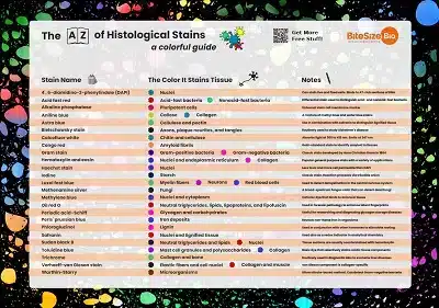
Obtain Your Free Histological Stains Poster
If you wish to transcend hematoxylin and eosin staining, why not obtain our free histological stains poster and brighten up your lab? It lists frequent stains, the colour they stain tissues, and a few helpful notes! Get your free copy right here.


