Experimental process
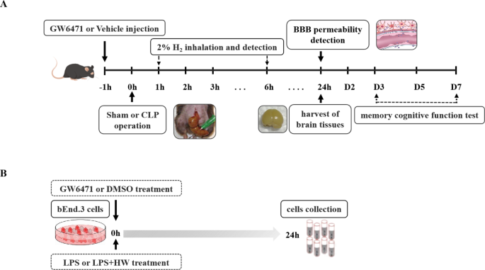
In vivo research (Fig. 1A): C57BL/6J male mice (6–8 w, 20–25 g) had been divided randomly into 4 teams: Sham, CLP, CLP + H2 and CLP + H2 + GW6471 teams. Sham or CLP surgical procedures had been carried out, and the H2 and GW6471 teams had been handled with H2 gasoline (for 60 min at 1 and 6 h postsurgery) and 20 mg/kg GW6471 (intraperitoneal injection at 1 h preoperation), respectively. Following the operations, the survival charges of all teams had been recorded, and a few mice had been euthanized 24 h postoperation with isoflurane, and mind tissue (cortex) was harvested. As well as, every group of mice was subjected to the concern conditioning take a look at (n = 5) and Y-maze take a look at (n = 3) (1–7 days after the process). Cortical tissues in mice had been used for Evans blue extravasation and mind water content material dedication. The cortex tissues had been used for P-gp/Abcb1, breast most cancers resistance protein (Bcrp/Abcb11), multidrug resistance-associated protein 2 (Mrp2/Abcc2), VE-cadherin, occludin and ZO-1 detection with WB. Mind slices from mice (n = 3) had been obtained for TUNEL staining and Nissl staining. The cortex tissues per group (n = 6) had been subjected to an enzyme-linked immunosorbent assay (ELISA) to judge inflammatory mediators (TNF-α, IL-6, IL-1β, and HMGB1).
Experimental design. (A) C57BL/6J mice (6–8 w, 20–25 g) had been subjected to sham or CLP operation. One hour after the injection of GW6471 or automobile, sham and CLP operations had been carried out. H2 or recent air was inhaled for 60 min ranging from 1 and 6 h postoperation with H2 focus detection. The mind tissue of various teams was obtained for checks 24 h after the sham or CLP process. Reminiscence cognitive operate checks had been carried out from 24 h to 7 days after the sham or CLP process. Completely different teams of mind tissues had been used for all of the examinations as described within the Supplies and Strategies. (B) Mouse bEnd.3 cells had been incubated with management + DMSO or GW6471 medium, management + HW + DMSO or GW6471 medium, LPS + DMSO or GW6471 medium, and LPS + HW + DMSO or GW6471 medium. The cells and tradition medium supernatant had been collected for testing 24 h after incubation. CLP, caecal ligation and puncture; LPS, lipopolysaccharide; GW6471, a PPARα inhibitor
In vitro research (Fig. 1B): bEnd.3 cells had been handled with the next circumstances: management + DMSO, LPS + DMSO, management + H2 + DMSO, LPS + H2 + DMSO, management + GW6471 (PPARα antagonist), LPS + GW6471, management + H2 + GW6471, and LPS + H2 + GW6471. To make clear the important results of PPARα, both 25 µM GW6471 (a PPARα antagonist) dissolved in DMSO (MCE, Tocris Bioscience, USA) or DMSO with out GW6471 was added to DMEM with out FBS, and the cells in all teams had been added to 1 µg/ml LPS (Cat# 4394, Sigma, USA) or the identical quantity of PBS/saline for twenty-four h. The LPS and management teams (DMSO or GW6471) had been cultured in regular DMEM with out FBS, and the management and LPS teams (+ H2 + DMSO or GW6471) had been developed in hydrogen-rich DMEM with out FBS for twenty-four h. In all teams, the cells had been harvested following the corresponding process at 24 h and centrifuged at 10,000 × g at 4 °C for 10 min. The liquid supernatant was obtained for Western blot evaluation.
bEnd.3 cell cultivation
Bought from the American Kind Tradition Assortment (ATCC), the bEnd.3 traces had been derived from brain-endothelial cells in mice and had been developed in Dulbecco’s modified Eagle’s medium (DMEM, Cat# PM150210, Procell, China) with 10% foetal bovine serum (FBS, Cat# 164210-50, Procell, China) and 1% streptomycin/penicillin answer (Cat# PB180120, Procell, China). Cultured in a 5% CO2 humid ambiance at 37 °C, the second generations had been utilized to the experiment when the bEnd.3 cells grew to 80% confluence in a Petri dish, and the subculture was administered.
Ethics assertion and animal preparation
Our analysis protocols had been carried out with the permission of the Animal Experimental Ethics Committee of Tianjin Medical College Common Hospital (No.IRB2020-DW-18) (Tianjin, China). Male C57BL/6J mice on this research had been offered by the Laboratory Animal Middle of the Navy Medical Science Academy (Beijing, China) and had been utilized in accordance with the Nationwide Institutes of Well being Information for Care and Use of Laboratory Animals. The managed circumstances had been appropriate for the mice (a humidity: 55 − 65%; temperature: 20-25 °C; 12/12-hour light-dark cycle) and given advert libitum water and meals.
Caecal ligation and puncture (CLP)
In our earlier research, we selected the CLP mannequin for the institution of sepsis in mice [30]. After adaptation for 7 days within the laboratory atmosphere, the mice had been anaesthetized with isoflurane and positioned within the supine place. After sterilizing the pores and skin within the stomach and making a 1 cm incision, 35% of the caecum was ligated after publicity. Subsequent, a 21-gauge needle was used to puncture twice the caecum, and sterile forceps had been utilized to push out roughly 0.3 mL of caecal content material. Lastly, the caecum was positioned again into the belly cavity, and the pores and skin and muscular tissues had been stitched. In distinction, the Sham group merely had the caecum uncovered after which had the belly cavity closed. After the operation, saline (1 mL) was subcutaneously utilized to the mice with lidocaine cream (Ziguang, Beijing, Cat# H20063466) to alleviate struggling. After the procedures, the animals had been positioned in a 20-25 °C room with a heating blanket.
Intraperitoneal injection of GW6471
GW6471 (Tocris Bioscience) was dissolved in DMSO to a focus of 12.5 mg/ml. A 20 mg/kg dose of GW6471 was intraperitoneally injected into mice 1 h earlier than the operation [31].
Hydrogen-rich medium
We made the hydrogen-rich medium (HM) with the strategy talked about in our earlier research [32, 33]. Briefly, with 0.4 MPa stress circumstances, the combination of H2 (1 l/min) and air (1 l/min) was dissolved in 5.6 mM glucose DMEM for at the least 4 h to acquire supersaturation (0.6 mM H2). A TF-1 gasoline move metre (Tokyo, Japan, Yutaka Engineering Corp.) was used to provide H2. A particular sealed aluminium vacuum package deal bag was used to retailer the ready HM at 4 °C beneath atmospheric circumstances. The medium have to be freshly ready each 7 days to maintain the H2 focus saturated.
Hydrogen inhalation
A field with two shops for H2 outflow and influx was utilized for the therapy of H2 for 1 h in mice at 1 and 6 h postsurgery. The combination of H2 and air was produced and infused into the field at a 4 l/min fee with a TF-1 gasoline move metre (Yutaka Engineering Corp., Tokyo, Japan). A detector (HY-ALERTA Handheld Detector, mannequin 500, H2 Scan, Valencia, Calif) was utilized to constantly monitor the focus of H2 maintained at 2% all through therapy within the field. Carbon dioxide was dissolved by baralyme within the field and discharged. The animals handled with out H2 had been positioned in the identical sort of field with room air [19].
Survival charges
As described beforehand, we recorded the survival charges of mice 7 days postoperation [18]. The experimental procedures had been carried out thrice.
Y-maze take a look at and contextual concern conditioning take a look at
The Y-maze was composed of A, B, and C arms with an angle of 120° from one another, which recorded the variety of alternations/occasions that each mouse strolled into all three arms in a row with out visiting one arm twice, i.e., for instance, the sample of ABC, BCA, or CAB. All mice had been positioned into the maze centre and given free exercise in all arms for 10 min. To file the variety of alternations and line crossings, we used the ANY-maze video monitoring system (Stoelting, USA), after which we analysed the mouse exercise to calculate the alternation proportion [30, 34].
Contextual concern conditioning take a look at: This take a look at is broadly utilized for evaluating reminiscence features [21, 35, 36]. The concern conditioning take a look at contained three levels: the a part of habituation, the a part of coaching, and the a part of the take a look at. Within the habituation part, mice had been put into the coaching context with free motion for 10 min. Within the coaching part, every group of mice was positioned in a concern chamber at some point earlier than modelling, acclimated for two min, after which given 20 s of a single-frequency sound sign (70 dB) coterminating with a foot shock (0.70 mA, 2 s). After an interval of 25 s, the auditory stimulus was performed for an additional 60 s coterminating with a second foot shock, marking the tip of 1 full cycle of coaching (105 s). Six cycles of coaching had been administered. The mice confirmed panic, escape, or rigidity after they heard the sound sign (no different motor behaviour besides respiratory) and squeal, soar and escape after they had been shocked, indicating the formation of concern reminiscence. The sham or CLP mannequin was established the day after concern reminiscence formation. Contextual concern reminiscence checks had been administered at 1 d, 2 d, 3 d, 5 d and seven d after sham surgical procedure or CLP. Through the take a look at interval, the mice in every group had been put into the concern field, which was the identical because the atmosphere through the coaching interval, however weren’t given the sound sign and electrical stimulation and got free motion for 300 s. The evaluation system of ANY-maze video was utilized to the time of rigidity file in every group through the coaching interval and the take a look at interval, and the proportion of time of rigidity was calculated. The mice had been considered freezing if there was no motion for two s (freezing time/300 s×100%= freezing time ratio).
Detection of inflammatory cytokines
Cortical tissues in all teams had been homogenized, centrifuged at 10,000 × g at 4 °C for 10 min and picked up with the supernatant. The degrees of IL-1β, TNF-α, IL-6 (Cat# RLB00; Cat# RTA00; Cat# R6000B; R&D Programs, Inc.) and HMGB1 (ARG81310; Arigo) had been measured by ELISA kits primarily based on the producer’s directions.
TUNEL staining
With the TUNEL assay, neuronal apoptosis was noticed at 24 h postsurgery. Fluorescein-dNTPs and fluorescein-dUTP compose the nucleotide-labelling combine (TUNEL reagent) and are used for in situ evaluation of apoptosis. The preparation of the TUNEL response combination requires the mixture of the nucleotide-labelling combine and the TUNEL enzyme. The cells with damaged DNA strands had been labelled by the response combination, offering for analysing and quantifying the extent of apoptosis on the single-cell stage. The cell nuclei had been stained with DAPI to turn into blue, and apoptotic cells had been stained inexperienced.
Nissl staining
Twenty-four hours after the operations, the animals’ brains had been obtained for the remark of the extent of mind damage. Mind tissues had been obtained after transcardial perfusion with 4% paraformaldehyde, postfixed for twenty-four h with formalin-free fixative after which embedded in paraffin. Following rehydration and deparaffinization with ethanol and dimethyl benzene, 10 μm mind sections had been used for Nissl staining. The injury within the cortex area was assessed with a microscope (CKX41, Tokyo, Japan).
EB extravasation within the blood‒mind barrier
As simply described, the BBB permeability was evaluated by the extravasation of EB dye [21]. At 24 h postoperation being anaesthetized, the mice had been handled with a 2.5 mL/kg dose of EB (the focus of two%, Cat# R31047, Shanghai Yuan Ye Bio-Know-how Co., Ltd, China) by way of the tail vein and noticed for two h. Then, all of the animals had been euthanized and transcardially perfused with a considerable amount of saline. The cortex of each mouse was collected, weighed and incubated for 48 h at 37 °C after homogenization in 1 mL formamide. After centrifugation, the optical density at 620 nm of the supernatant liquid was calculated with a microplate reader. Primarily based on a linear customary curve, a big amount of EB was quantified (µg/g moist weight) after which proven because the relative quantity [32].
Mind water content material (WC)
The animals had been euthanized at 24 h to reap complete mind tissues after surgical procedure. The water content material within the mind was assessed with a dry‒moist methodology as described in earlier analysis [21]. After being weighed immediately, the mind tissues had been dried at 100 °C in an oven for twenty-four h, after which the moist weight and dry weight had been obtained. Primarily based on the next components, WC = 100% × [wet weight/dry weight].
Western blot (WB)
WB was used to judge the expression of PPARα, MRP2, P-gp, BCRP, ZO-1, VE-cadherin and occludin. Twenty-four hours after the operation, the cortex tissue from every group was collected and weighed. The protease inhibitor and PBS had been added to every pattern, and after centrifugation at 4 °C for 15 min at 15,000 × g, we obtained the supernatants after which quantified the collected protein concentrations. The samples had been added to the loading buffer (Beijing Solarbio Science & Know-how Co., Ltd. ), boiled and denatured. The proteins had been separated by sodium dodecyl sulfate‒polyacrylamide gel electrophoresis (SDS‒PAGE) and electrotransferred onto polyvinylidene fluoride membranes (Millipore, Germany). Subsequent, the membranes had been soaked in Tris-buffered saline with Tween (TBST) with 5% nonfat milk for two h and incubated within the following main antibodies at 4 °C in a single day: PPARα, MRP2, P-gp, BCRP (1:1000, Cat# ab61182; ab203397; ab170904; ab130244, Abcam, Britain), ZO-1 (1:1000, Cat#AF5145, Affinity, USA), VE-cadherin (1:500, Cat# AF6265, Affinity, USA), and occludin (1:1000, Cat# DF7504, Affinity, USA). After washing with TBST, the membrane was soaked in TBST containing goat anti-rabbit (1:5000, Cat# 31,466, Invitrogen, USA) or anti-mouse antibodies (1:5000, Cat# 31,430, Invitrogen, USA) for 1 h at room temperature. The nitrocellulose membrane was immersed in electrochemiluminescence (ECL) reagent. The protein bands had been analysed by ImageJ. The degrees of the goal proteins had been standardized to β-actin or GAPDH.
Statistical evaluation
GraphPad Prism 8.0 and SPSS 21.0 software program had been used to analyse the information. The Shapiro‒Wilk take a look at and KS normality take a look at had been used to find out whether or not a pattern got here from a standard distribution. The survival charges had been assessed as percentages after which analysed by the log‑rank (Mantel‒Cox) take a look at. Knowledge from the behavioural take a look at and H2 focus detection had been analysed utilizing two-way evaluation of variance (ANOVA) with Tukey’s a number of comparisons take a look at, and the ELISA, Western blot, Evans blue extravasation and mind water content material knowledge had been analysed by one-way ANOVA with Tukey’s a number of comparisons take a look at. Quantifiable knowledge are expressed because the imply ± customary deviation (SD). P < 0.05 was acknowledged as statistically important in all checks.


