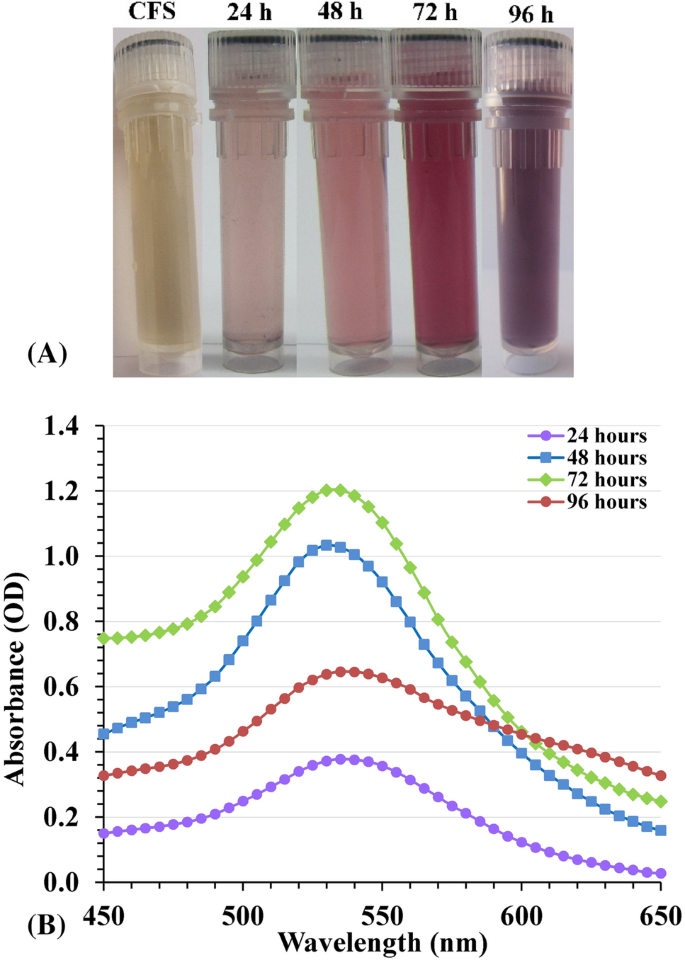
There’s a rising demand to develop eco-friendly, non-toxic, cost-effective and dependable routes of gold nanoparticles synthesis. The biosynthesis of AuNPs has been completed utilizing the cell-free supernatant of Streptomyces flavolimosus. The cell-free supernatant was handled with aqueous answer of HAuCl4. The response combination was incubated in an incubator shaker at nighttime situation to keep away from photolytic response at 37 °C for twenty-four–72 h. After the incubation time, the discount of HAuCl4 to AuNPs was monitored visually and the change within the coloration of the response combination from yellow, violet to purple or darkish purple was noticed, indicating the discount of gold chloride and formation of AuNPs (Fig. 1A). In distinction, no color change was noticed in a management combination when the cell-free supernatant was incubated beneath the identical circumstances with out the addition of aqueous HAuCl4 answer.
The mechanism of the biogenic course of isn’t totally understood. The biosynthesis technique of AuNPs takes place in two steps: on the first, the gold ions (Gold (III) chloride, historically referred to as auric chloride, HAuCl4, chloroauric acid, Au3+) are reworked to AuNPs (Au0) in presence of decreasing brokers, and adopted by the coating of synthesized AuNPs. In the course of the inexperienced synthesis processes, all kinds of natural in addition to organic molecules (enzymes, phenols, sugars, and many others.) current in organic samples can take part each within the gold ions discount and within the ample capping and stabilization of the ready nanoparticles13,25. It’s believed that enzymatic synthesis is among the most effective attainable strategies for producing AuNPs. The enzyme nitrate reductase was discovered to catalyze and play an important position within the technique of gold ions discount13.
UV–Seen spectral evaluation
AuNPs biosynthesis was explored utilizing UV–Seen spectroscopy within the spectral area 450–650 nm, with the development of the interval. Characterization of AuNPs begins with a color change based mostly on the floor Plasmon resonance precept (SPR). SPR is the optical property that’s distinctive to metallic nanoparticles. The steel floor carry free electrons within the conduction band and the nuclei are positively charged. SPR is attributable to the oscillations of electrons within the conduction band near the nanoparticles’ floor. The precise oscillations modes of electrons restricted by the particles measurement and form. Consequently, metallic nanoparticles exhibit distinctive optical absorption spectra within the ultraviolet–seen vary. When the dimensions of the particles will increase, a coloration change takes place; within the case of gold, this modification goes from a deep purple to a purple coloration. The SPR is liable for the variations in coloration noticed throughout AuNPs biosynthesis and UV–Seen spectroscopy is used to find out the absorbance of the assorted coloration modifications. The optical properties of AuNPs–from gentle pink to darkish red-are managed by their morphology, which incorporates their measurement, form, the diploma to which they mixture and structural traits of those nanoparticles26.
To determine the impact of the cell-free supernatant on AuNPs biosynthesis, the absorbance spectrum of the cell-free supernatant was carried out (Supplementary Fig. S1). SPR spectrum of AuNPs synthesized utilizing Streptomyces flavolimosus cell-free supernatant confirmed the presence of a definite absorption peak with most absorbance wavelength positioned at 530–535 nm (Fig. 1B). It’s similar to the SPR spectra of AuNPs. The outcomes point out that full discount of the HAuCl4 ions happens after almost 3 days of the response incubation time. Ahmad et al.27 reported that the SPR band of AuNPs answer’s staying at 530 nm throughout the period of the response interval proves that there is no such thing as a signal of aggregation and that the particles are dispersed within the aqueous answer. Full discount of the HAuCl4 ions takes roughly 120 h, demonstrating that this can be a very sluggish course of27. Doan et al.28 noticed that the presence of a single absorption peak exhibiting most absorbance at round 535 nm is attributable to the AuNPs SPR band, and the focus of gold ions have a major impression on the AuNPs biosynthesis. The rise in UV–Vis absorbance was attributable to the rise in gold ion focus. Kalabegishvili et al.29 reported that, the synthesis of AuNPs was attributable to the addition of Streptomyces glaucus 71MD biomass to chloroauric acid and the utmost absorption peak indicating the biosynthesis of gold nanoparticles was noticed at 530 nm. However, Khadivi Derakhshan et al.30 acknowledged that UV seen absorption spectrum of AuNPs produced with Streptomyces griseus cells exhibited a outstanding and broad peak at 540 nm.
After three days of the response time, a lower within the absorbance was observed within the depth of the SPR band at 96 h. Additionally, the absorbance spectrum change into broadened at 96 h indicating the opportunity of AuNPs agglomeration. Sobczak-Kupiec et al.31 reported that shifting in addition to broadening of the floor plasmon band (SPB) of gold nanoparticles, reveals the formation of gold nanoparticles with almost fixed measurement distribution. The absorbance shift signifies the interlayer aggregation of gold nanoparticles. On very long time response, the initially fashioned particle serves as a nucleation website and because of this layer by deposition of gold takes place ensuing within the enhance in measurement31. However, Gupta et al.32 noticed that with rising of time, the AuNPs agglomerate and fuse to turn into bigger crystals, and thus large-sized AuNPs are produced.
Transmission electron microscopy (TEM) evaluation
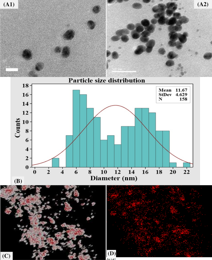
The morphology, microstructures, and measurement of the biosynthesized AuNPs have been decided by TEM evaluation. TEM is an efficient device for investigating nanoparticles and is well known for inspecting morphological traits, similar to form, measurement, and floor space33. Determine 2A demonstrates that TEM investigation revealed the formation of well-dispersed spherical AuNPs with sizes starting from 4 to twenty nm. Whereas Fig. 2B shows a plot of the histogram of particle measurement distribution that was calculated based mostly on an evaluation of 158 particles with imply measurement distribution of 11.67 nm.
Protocols for the organic synthesis of gold nanoparticles present a clear and eco-friendly methodology for the manufacturing of nanoparticles with a broad vary of sizes, shapes, physico-chemical and organic properties34. Huang et al.35 reported that attribute size-distribution and TEM photographs of the AuNPs have disclosed spherical formed AuNPs with a broad spectrum of particle measurement with the dimensions starting from 5 to 50 nm. Moreover, Cai et al.36 reported that TEM picture for AuNPs biosynthesized by Magnetospirillum gryphiswaldense MSR-1 present spherical nanoparticles, with an irregular measurement distribution within the vary of 5–40 nm. Rajeshkumar37 reported that TEM investigation for AuNPs biosynthesized by marine micro organism Enterococcus sp. demonstrated the formation of spherical formed AuNPs with measurement starting from 6 to 13 nm.
Mapping evaluation of AuNPs was carried out to analyze the distribution and the composition of biosynthesized AuNPs. TEM elemental mapping evaluation outcomes illustrate the entire distribution of AuNPs and its elements (Au) (Fig. 2C).
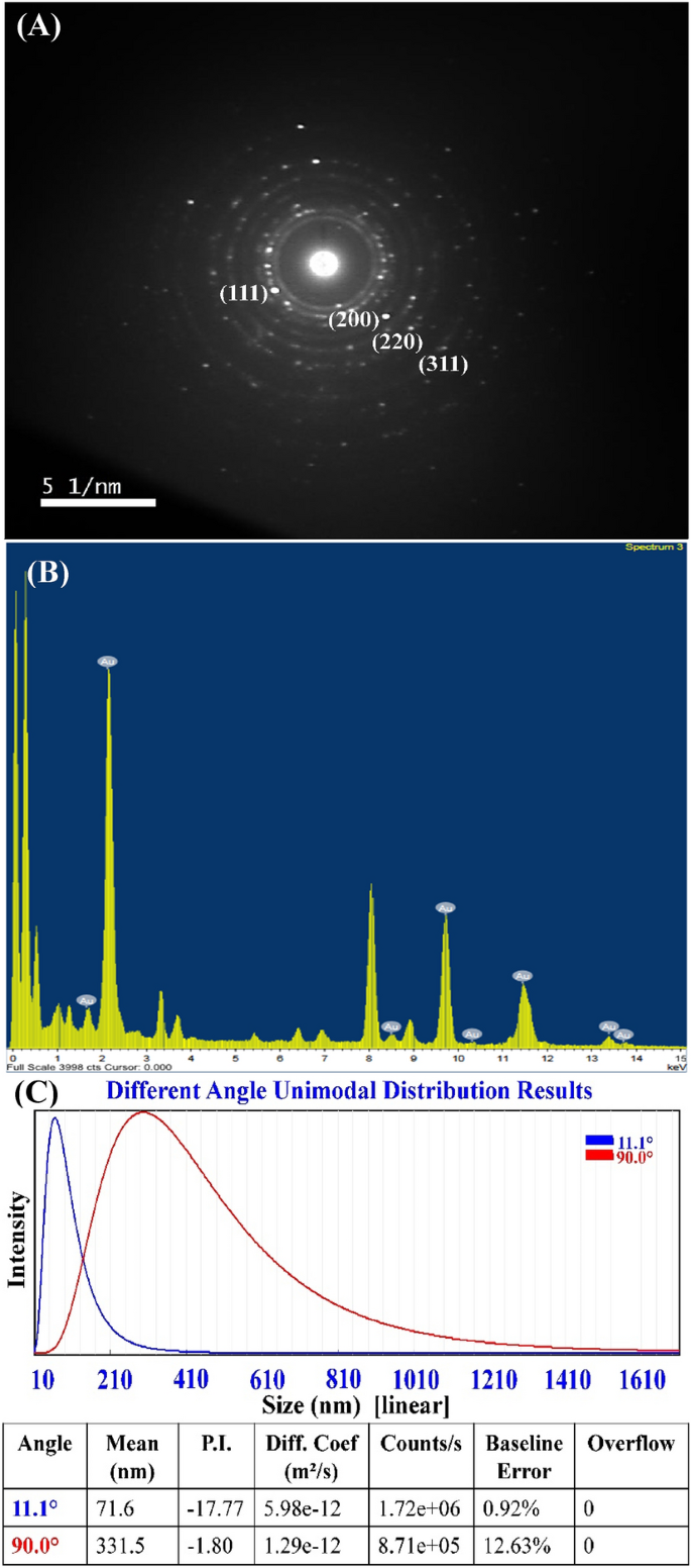
To verify AuNPs formation and its crystalline construction, SAED (chosen space electron diffraction sample) evaluation was carried out (Fig. 3A). SAED patterns obtained by TEM have been used to judge the crystallinity of the AuNPs38. AuNPs crystalline construction was demonstrated by the intense round well-separated diffraction rings associated to the next 4 lattice planes: (111), (200), (220), and (311), which indicated the fcc (face-centered cubic) construction of the gold38.
Determine 3B reveals customary EDX spectrum of the biosynthesized AuNPs. EDX by TEM is an analytical methodology used to find out the fundamental composition of a pattern33. The existence of gold within the produced AuNPs was confirmed by EDX evaluation39.
Particle measurement evaluation
Particle Measurement Analyzer (PSA) was used on this investigation to evaluate the particle measurement distribution of an AuNPs suspension. The particle measurement distribution was decided at ambient temperature over a variety of wavelengths from 1 to 760 nm. As proven in Fig. 3C, a particle measurement analyzer measurement of a pattern of AuNPs revealed a slim and sharp peak at 71.6 nm diameter at θ = 11.1° and 331.5 nm at θ = 90°. The sizes that have been measured by the particle measurement analyzer can solely be used as a relative worth, and can’t be in comparison with these decided by electron microscopy40. Electron microscopy is ready to decide the form and floor construction of the particles and their geometric dimensions by measuring the width of particular person particles from the picture41,42. Imaging was favored due to its high-resolution particle’s visualization and negligible impression of artifacts on measurement dedication42.
Zeta potential (ζ) measurement
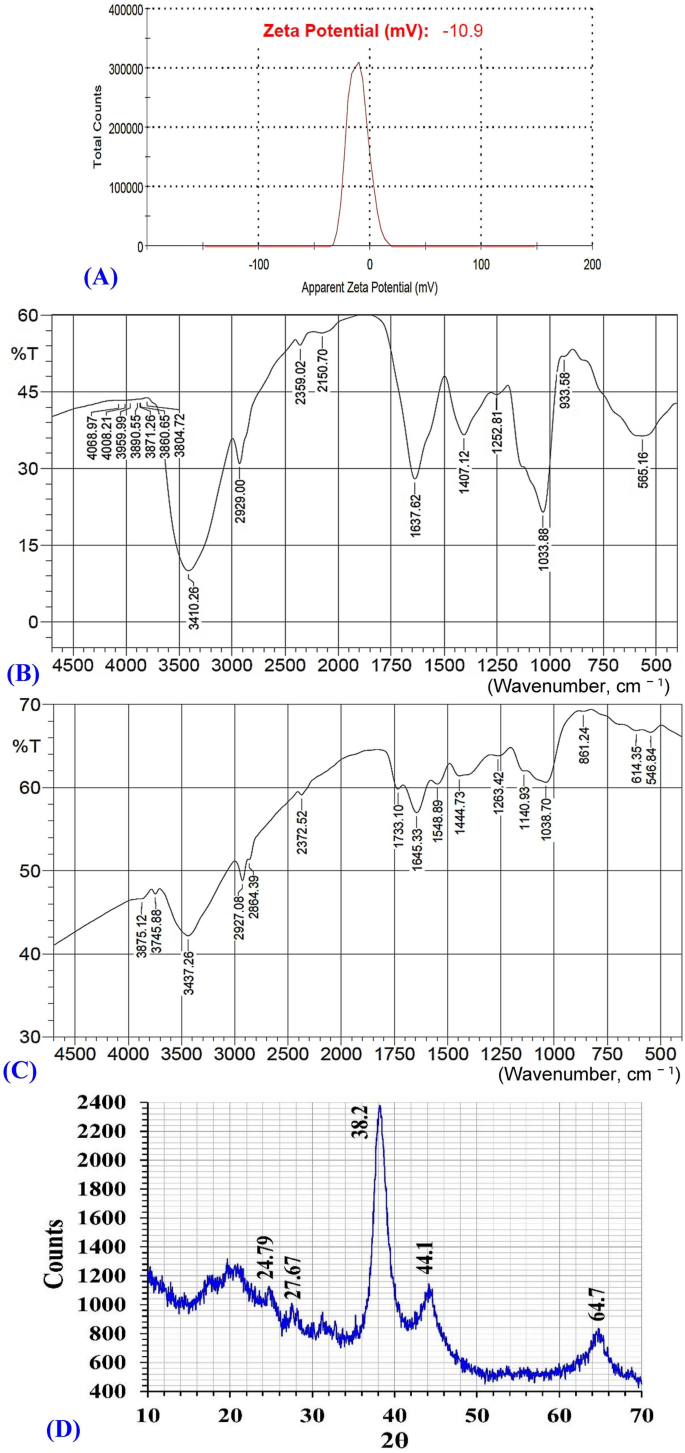
The web cost of nanoparticles is a key attribute that influences their stability and dispersion33. Subsequently, the zeta potential (ζ) values have been used to find out the soundness and the floor cost of the biosynthesized AuNPs. Zeta potential is measured relying on the rate and path of particles in a well-defined electrical subject43. Bhambure et al.44 reported that zeta potential defines the particle’s repelling forces, which forestall aggregation and is liable for stabilization of the synthesized AuNPs within the answer. Determine 4A demonstrates that the biosynthesized AuNPs confirmed zeta potential worth of − 10.9 mV. The adverse zeta potential worth signifies that AuNPs are surrounded by negatively charged natural molecules, which reduces the repulsion between the AuNPs, prevents their aggregation, and subsequently will increase their stability45.
The zeta potential worth was discovered to be − 9.8 mV46. The synthesized AuNPs confirmed adverse zeta potential worth of − 2.26 mV45. Two various kinds of AuNPs synthesized from Hashish sativa confirmed zeta potential values of − 12.3 mV and − 20.6 mV46. The excessive values of zeta potential imply that AuNPs are extremely secure because of the presence of excessive floor cost that forestall aggregation and guarantee redispersion as a consequence of repulsive electrical forces. At values of zeta potential, aggregation could type47. As a normal rule, zeta potential worth ≥ 30 mV is taken into account good and wonderful stability47. Values of zeta potential ≥ ± 30 mV signifies monodisperse formulations with out aggregates48, whereas zeta potential worth ~ ± 20 mV is vulnerable to have solely short-term stability, and zeta potential worth < 5 mV tends to mixture quickly47.
Since zeta potential is influenced by temperature, solvent viscosity, pH, ionic power, and floor traits, even minor parameter variations can considerably change its absolute worth49. Furthermore, altering the focus of gold salt used for the synthesis response, pH, and temperature can even present management over the dimensions and geometry of AuNPs50.
The secure nanoparticles are stabilized in presence of stabilizing brokers. Varied research have reported that proteins, terpenoids, phenolic compounds, and nicotinamide adenine dinucleotide may act as capping and stabilizing brokers throughout AuNPs biosynthesis50. In response to the findings of Muthuvel et al.43, the capping molecules which can be discovered on the floor of AuNPs are predominantly made up of teams which have a adverse cost. The varied useful teams similar to carboxylic, polyphenol, alcohol, alkene, main amine, and surfactant presence in proteins could management the form and measurement of nanoparticles with the change of salt precursors, pH, and temperature25. The stabilization of NPs finished through the use of varied capping brokers could be divided into three stabilization processes, together with steric stabilization, electrostatic stabilization, and unification of steric and electrostatic stabilization25,51.
Fourier reworked infrared evaluation (FTIR)
Inexperienced synthesis of AuNPs was achieved utilizing the cell-free supernatant of Streptomyces flavolimosus as a biocatalyst for discount of HAuCl4 and capping/stabilizing of the AuNPs. The FTIR evaluation was carried out to find out the biomolecules liable for decreasing, capping, and stabilizing brokers of AuNPs. FTIR evaluation was carried out with a purpose to determine the useful teams located on the AuNPs’ floor52 or figuring out the possible biomolecules liable for the discount of Au+ ions by the cell-free supernatant53.
FTIR spectrum of the bio-synthesized AuNPs was analyzed and in contrast with the FTIR spectrum of the cell-free supernatant of Streptomyces flavolimosus (Fig. 4B,C). The FTIR spectrum of the cell-free supernatant of Streptomyces flavolimosus (Fig. 4B) reveals absorption peaks at 3745, 3437, 2927, 2372, 1733, 1645, 1548, 1444, 1263, 1140, 1038, 861, 614 and 546 cm−1. Vital shifts of peaks within the spectrum of AuNPs from peaks within the spectrum of the cell-free supernatant of Streptomyces flavolimosus (Fig. 4C) point out a major position of varied operate teams within the cell-free supernatant of Streptomyces flavolimosus within the discount, capping, and stabilizing AuNPs. The FTIR spectrum of AuNPs (Fig. 4C) reveals distinct absorption peaks at 3410, 2929, 2359, 2150, 1637, 1407, 1252, 1033, 933 and 565 cm−1.
Hydroxyl (OH) stretching peak at wavelength 3437 cm−1 discovered within the spectrum of cell-free supernatant of Streptomyces flavolimosus shifted to 3410 cm−1 which is attributed to –OH stretching54 on the formation of AuNPs. Furthermore, the absorption equivalent to C–H stretching vibration was noticed at 2927 cm−1 discovered within the spectrum of cell-free supernatant of Streptomyces flavolimosus shifted to 2929 cm−1 which is related to the C–H stretching of alkanes55 on the formation of AuNPs. The C=O or the N–H stretching vibrations have been considered liable for the height that was seen at roughly 2359 cm−156. The band positioned at 2150 cm−1 could possibly be assigned to C=O57, it may be attributed to amine hydrochloride stretching vibration and the presence of NH3+58.
The peaks at 1645 cm−1 discovered within the spectrum of cell-free supernatant of Streptomyces flavolimosus corresponds to amide I as a consequence of C=O stretching of α-helix proteins59 shifted on the formation of AuNPs to the absorption peak located at 1637 cm−1 assigned to the amide group I, and it’s attributable to the stretching vibrations of carbonyl which can be current in proteins amide linkage60. However, the presence of peak at 1548 cm−1 within the FTIR spectrum of cell-free supernatant of Streptomyces flavolimosus is because of the N–H bending vibrations of the protein amide II group. The height at 1548 cm−1 disappeared within the FTIR spectrum after AuNPs biosynthesis, reveals that this teams is concerned within the AuNPs biosynthesis by the tradition filtrate of Streptomyces flavolimosus.
The C–C stretching of fragrant molecules is represented by the bands at 1407 cm−155 and C–O stretching vibrations61. Moreover, the height of the C–N stretching vibrations seen within the spectral vary of 1250 to 1000 cm−162. The band noticed at 1252 cm−1 is assigned to the C–C stretching vibrations63. The absorption peaks at 1038 cm−1 discovered within the spectrum of cell-free supernatant of Streptomyces flavolimosus could possibly be because of the stretching vibration of the C–O teams within the monosaccharides shifted on the formation of AuNPs to the absorption peaks at 1033 cm−1 corresponds to the C–O stretching vibrations which can be current in varied biocomponents64. The height at 933 cm−1, is indicative of hydrogen bridging these outcomes from the technology of carboxylic acid dimers65.
X-ray diffraction sample (XRD)
XRD sample of AuNPs biosynthesized through the use of the cell-free supernatant of Streptomyces flavolimosus was carried out (Fig. 4D). The peaks of AuNPs have been discovered at 2θ = 24.79°, 27.67°, 38.2°, 44.1°, 64.7° (Fig. 4D), within the vary of 20°–70°. The standard peaks of AuNPs have been positioned at 2θ = 38.1°, 44.3°, 64.6°, 77.6°, 81.7°, 98.2°, 111.0° and 115.2°66. Doan et al.28 acknowledged that the utmost peak of diffraction for AuNPs occurring at 2θ angle of roughly 38.2° indicated that the crystals have a favoured progress orientation within the airplane (111) and the end result obtained clearly proves that the AuNPs fashioned have been crystalline in nature67.
Statistical optimization of AuNPs biosynthesis utilizing the cell-free supernatant of Streptomyces flavolimosus pressure utilizing central composite design (CCD)
Many components, together with incubation time, temperature, pH, and HAuCl4 focus, can affect the dimensions, distribution, and amount of gold nanoparticles which can be synthesized. Within the present research, the results of 4 components on AuNPs biosynthesis (as a response) have been explored. These 4 variables have been the HAuCl4 focus, the preliminary pH degree, temperature, and the incubation interval.
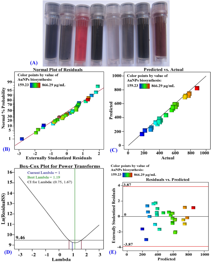
The bioprocess variables affecting the AuNPs biosynthesis have been optimized through the use of the CCD with a purpose to maximize the AuNPs biosynthesis and to analyze the quadratic, interplay, and particular person results of the method variables on the biosynthesis of AuNPs through the use of the cell-free supernatant of Streptomyces flavolimosus. With a purpose to decide the optimum values for the variables of curiosity, a CCD consisting of thirty separate trials was utilized. Desk 1 illustrates the design matrix containing the 4 investigated variables, their precise and coded ranges, in addition to the experimental and predicted biosynthesized AuNPs values and their residual values. The outcomes that have been obtained from the CCD experiments for the biosynthesized AuNPs utilizing the cell-free supernatant of Streptomyces flavolimosus reveal that there are vital coloration modifications mirrored by the biosynthesized AuNPs as affected by the degrees of various unbiased variables throughout the optimization course of (Fig. 5A).
(A) Images of the AuNPs options present coloration modifications as affected by the degrees of various unbiased variables throughout the optimization course of. (B) NPP of internally studentized residuals, (C) plot of predicted versus precise, (D) Field–Cox plot for energy transforms and (E) plot of the residuals versus predicted values of the biosynthesized AuNPs utilizing the cell-free supernatant of Streptomyces flavolimosus.
In response to the info that was collected, the values of the biosynthesized AuNPs ranged from a 159.23 to 866.29 µg/mL. The best worth of biosynthesized AuNPs (866.29 µg/mL) was obtained within the trial quantity 2 the place the gold focus was 1000 µg/mL, the pH was 6, the temperature was 35 °C, and the incubation time was 4 days. In distinction, the bottom worth of biosynthesized AuNPs (159.23 µg/mL) was produced in run quantity 30 with gold focus of 600 µg/mL, pH of 4, temperature of 35 °C, and incubation time of 4 days.
A number of regression evaluation and the evaluation of variance (ANOVA)
Desk 2 shows the outcomes of the evaluation of variance (ANOVA) carried out on the experimental knowledge of CCD for the biosynthesis of AuNPs utilizing the cell-free supernatant of Streptomyces flavolimosus. The mannequin’s validity was assessed utilizing the info recorded in Desk 2, together with adjusted R2 worth, R2 worth, predicted R2 worth, P-value, lack of match and F-value and the coefficient estimate values. As well as, the linear, interplay, and quadratic impacts of the 4 chosen course of variables have been evaluated15.
The dedication coefficient (R2) was utilized to measure the adequacy of the mannequin because it quantifies the extent of response worth variability that could possibly be attributable to the experimental parameters. R2 values are all the time lower than or equal to 1, and when they’re nearer to 1, it signifies that the mannequin is extra strong and may estimate the response extra exactly. The mannequin that had an R2 worth that was greater than 0.9 was regarded to have a really excessive correlation68. Within the present research, the R2 worth of the mannequin used for the biosynthesized AuNPs is 0.9885 (Desk 2), reflecting that 98.85% of variance within the biosynthesized AuNPs is attributable to the unbiased variables and simply 1.15% of the full variance within the biosynthesized AuNPs isn’t defined by the unbiased components. R2 worth mirrored an excellent match between the experimental and predicted values of the biosynthesized AuNPs. However, the regression mannequin used for AuNPs biosynthesis has an adjusted dedication coefficient (Adjusted R2) equal to 0.9778 (Desk 2). Additionally, the excessive worth of predicted R2 (0.9392) was in an affordable settlement with adjusted R2 worth which signifies that the mannequin is sweet for predicting AuNPs biosynthesis to new experiments. The worth of predicted R2 (0.9392) was in shut settlement with the adjusted R2 worth (0.9778). This demonstrated that there’s a excessive compatibility between the noticed and predicted values of the response14.
The worth of enough precision assesses the signal-to-noise ratio. A signal-to-noise ratio increased than 4 is fascinating and reflective of the mannequin’s precision69. Within the current research, the mannequin’s enough precision worth was 35.34, indicating enough design house for optimizing the biosynthesis of AuNPs at completely different values of the examined parameters.
As well as, the estimated coefficient demonstrated constructive or adverse results on the biosynthesis of AuNPs. There are two sorts of interactions that may happen between two variables: antagonism (a adverse coefficient) and synergism (a constructive coefficient). If the estimated impact worth is excessive, no matter whether or not it’s constructive or adverse, this means that the variable has a considerable impression on the response. However, if the impact is near zero, this means that the variable has little or no affect on the response. If the coefficient of a examined variable has a constructive signal, it signifies that yield will increase because the variable’s worth will increase. Conversely, a adverse signal signifies that yield will increase on the low worth of the variable70. If the coefficient for interactions between two variables is constructive, this means that the interactions between the variables have a synergistic impact on the biosynthesis of AuNPs. The outcomes indicated that the interplay results between X1 X2, X1 X3, X2 X4, X3 X4 (Desk 2) enhance AuNPs biosynthesis. Nevertheless, the presence of a adverse coefficient signifies that the interactions between two components have an antagonistic impact and reduce AuNPs biosynthesis. The outcomes indicated that the interplay results between X1 X4, X2 X3 lower AuNPs biosynthesis.
F-values and likelihood values (P-values) (Desk 2) have been additionally estimated to find out the importance of every coefficient, which is crucial for assessing the components significance and for deciphering the sample of their mutual interactions. Furthermore, course of components with P-values ≤ 0.05 have been thought of to have vital results on the response70. The outcomes of the Fisher’s F-test (92.17), together with a really low P-value (< 0.0001), are offered in Desk 2, and they reveal that the mannequin is very vital. Based mostly on the P-values of the coefficient, it may be concluded that linear results of temperature (X1), incubation instances in days (X2), HAuCl4 focus ((upmu)g/mL) (X3) and preliminary pH degree (X4) are vital for AuNPs biosynthesis utilizing the cell-free supernatant of Streptomyces flavolimosus. X1, X2, X3 and X4 had F-values of 13.71, 52.62, 797.39, and 76.88, and P-values of 0.0021, < 0.0001, < 0.0001, and < 0.0001; respectively. This extremely vital that the 4 variables contribute as limiting variables. This means that the 4 variables contribute as limiting variables. Minor variations of their ranges will have an effect on AuNPs biosynthesis utilizing Streptomyces flavolimosus cell-free supernatant. Moreover, the P-values of the coefficient revealed that every one interactions between the 4 examined components have been vital for AuNPs biosynthesis utilizing Streptomyces flavolimosus cell-free supernatant.
The findings of the match abstract which can be supplied in Desk 3 have been used to find out which of the linear, 2FI, and quadratic fashions was probably the most acceptable mannequin for the AuNPs biosynthesis utilizing the cell-free supernatant of Streptomyces flavolimosus. The quadratic mannequin is an acceptable mannequin for the biosynthesis of AuNPs because the lack of match is non-significant (P-value is 0.067 and the F-value is 4.08). Moreover, the R2 of 0.9885, the adjusted R2 worth (0.9778), in addition to the anticipated R2 worth (0.9392), are larger within the quadratic mannequin compared to the values obtained in different fashions (Desk 3). Quadratic mannequin abstract statistics for AuNPs biosynthesis confirmed a minimal customary deviation of 26.47. The anticipated residual sum of squares (PRESS) demonstrates the validity of the mannequin. If the PRESS statistic is low, the info factors are well-fit by the mannequin. The quadratic mannequin achieves a minimal PRESS worth of 55,565.78.
Through the use of a number of regression evaluation to the experimental outcomes, the next second-order polynomial equation was used to find out the anticipated AuNPs biosynthesis (Y) that correspond to the optimum ranges of preliminary pH, HAuCl4 focus, incubation interval, and temperature:
$${textual content{Y}} = { 575}.{17 } + { 2}0{textual content{ X}}_{{1}} {-}{ 39}.{textual content{19 X}}_{{2}} + { 152}.{textual content{55 X}}_{{3}} + { 47}.{textual content{37 X}}_{{4}} + {17}.{textual content{98 X}}_{{1}} {textual content{X}}_{{2}} + { 43}.{textual content{36 X}}_{{1}} {textual content{X}}_{{3}} {-}{19}.{textual content{82 X}}_{{1}} {textual content{X}}_{{4}} {-}{ 32}.{textual content{95 X}}_{{2}} {textual content{X}}_{{3}} + { 37}.{textual content{35 X}}_{{2}} {textual content{X}}_{{4}} + { 25}.{textual content{46 X}}_{{3}} {textual content{X}}_{{4}} {-}{1}.{textual content{8 X}}_{{1}}^{2} , {-}{1}0.{textual content{37 X}}_{{2}}^{2} , {-}{8}.{textual content{85 X}}_{{3}}^{2} , {-}{74}.{textual content{24 X}}_{{4}}^{2} .$$
Y is the anticipated AuNPs biosynthesis, X1 (temperature), X2 (incubation interval), X3 (HAuCl4 focus) and X4 (preliminary pH).
The mannequin adequacy
The traditional likelihood plot (NPP) is a metric that verifies the health of a mannequin by indicating whether or not or not the residuals comply with a standard distribution23. Residuals are variations between theoretical predictions and experimental findings; A low worth means the mannequin is enough71. Determine 5B reveals the NPP, which reveals that the residuals happen alongside the diagonal straight line of AuNPs biosynthesis. This illustrates that the anticipated knowledge well-fit with the experimental outcomes, which verifies the accuracy of the mannequin58.
Determine 5C compares the experimental outcomes with predicted values for the biosynthesis of AuNPs through the use of the cell-free supernatant of Streptomyces flavolimosus. It demonstrates that the entire factors are positioned very near the prediction line, which means that the mannequin adequately matches the experimental knowledge71.
As well as, Fig. 5D reveals Field–Cox plot of mannequin transformation. It demonstrates that the purple strains characterize the high and low 95% confidence ranges equivalent to the very best λ worth (0.75 and 1.67; respectively). The blue line represents the present transformation (λ = 1) whereas the inexperienced line represents the most effective worth of λ (λ = 0.73). Because the blue line lies between the 2 purple strains, the mannequin is within the optimum zone. This suggests that no knowledge transformation is required, and the mannequin is an efficient match for the obtained experimental outcomes58.
Determine 5E presents the externally studentized residuals towards the values that have been predicted for the AuNPs biosynthesis utilizing the cell-free supernatant of Streptomyces flavolimosus. The graph revealed that the residuals are distributed randomly across the zero line, which means that the experimental outcomes have a continuing small variance72.
Three dimensional (3D) plots
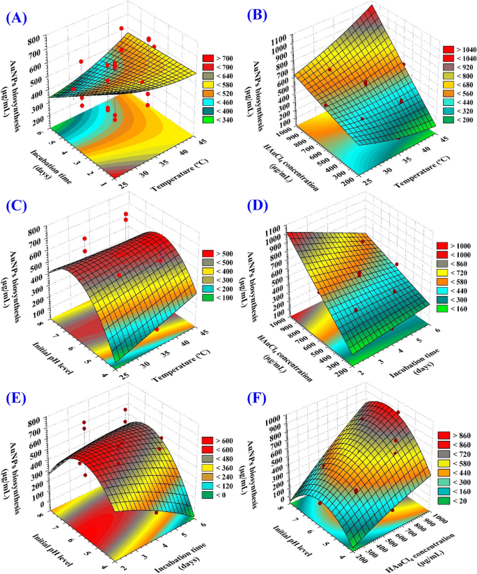
Determine 6A–F depicts the connection between the biosynthesized AuNPs and the mutual interactions between studied variables to find out the optimum circumstances for AuNPs biosynthesis utilizing the cell-free supernatant of Streptomyces flavolimosus. For the pair-wise mixtures of the 4 variables, 3D plots (X1 X2, X1 X3, X1 X4, X2 X3, X2 X4, X3 X4) have been generated by plotting AuNPs biosynthesis on Z-axis towards the X and Y axes for 2 unbiased variables whereas preserving the values of the opposite two variables at their heart factors.
The 3D plot (Fig. 6A) demonstrating the mutual interactions of the temperature (X1) and the incubation time (X2) on the biosynthesized AuNPs, whereas HAuCl4 focus (X3), and preliminary pH degree (X4) have been maintained at their central ranges. Determine 6A demonstrates that decreased ranges of each temperature (X1) and incubation time (X2) assist the very best AuNPs manufacturing, whereas the AuNPs biosynthesis lower regularly with rising temperature or incubation time.
The impact of temperature on AuNPs biosynthesis
Within the present research, the optimum temperature for max gold nanoparticles biosynthesis was 35 °C. Kulkarni et al.73 noticed that the nanoparticles formation fee and, consequently, the nanoparticles measurement could possibly be managed by adjusting parameters similar to gold focus, temperature, pH, and publicity time to HAuCl4. Camas et al.74 confirmed that the optimum temperature for max gold nanoparticles biosynthesis was 35 °C and gold nanoparticles biosynthesis is negatively impacted by a rise in temperature. The optimum temperature for max gold nanoparticles biosynthesis by Streptomyces sp. ERI-3 supernatant was 30 °C75.
The impact of incubation time on AuNPs biosynthesis
Within the present research, the optimum incubation time for max gold nanoparticles biosynthesis was 4 days. Zonooz et al.75 demonstrated that increased yield of gold nanoparticles by Streptomyces sp. ERI-3 supernatant was produced after 96 h of incubation. However, Camas et al.74 demonstrated that the utmost gold nanoparticles biosynthesis by Citricoccus sp. K1D109 was produced after 24 h of incubation, and that the biosynthesis decreases because the interplay time will increase.
Determine 6B describes the connection between the biosynthesized AuNPs and the mutual interactions between the temperature (X1) and the HAuCl4 focus (X3), whereas incubation time (X2) and preliminary pH degree (X4) have been maintained at their central ranges. It may be seen that the biosynthesized AuNPs elevated regularly by rising the temperature to 35 °C and the HAuCl4 focus close to to (1000 µg/mL).
The impact of HAuCl4 focus on AuNPs biosynthesis
With rising gold chloride focus, the biosynthesis of AuNPs was seen to extend. It could possibly be attributable to the excessive focus of gold chloride, the place extra Au+3 ions have been obtainable for discount to AuNPs76. Zonooz et al.75 acknowledged that the excessive titer of AuNPs biosynthesis by Streptomyces sp. ERI-3 supernatant (1450 a.u.) was achieved with an answer of three mM HAuCl4. Nevertheless, the synthesis of AuNPs was decreased at concentrations of three.5 and 4 mM, principally in all probability due to the toxicity of steel ions on elements concerned within the synthesis of AuNPs.
Determine 6C highlights the connection between the biosynthesized AuNPs and the mutual interactions between the temperature (X1) and preliminary pH degree (X4), whereas incubation time (X2) and HAuCl4 focus (X3) have been maintained at their central ranges. The biosynthesized AuNPs worth elevated till the optimum pH (at 6) was reached, after which decreased when the pH degree rose above that. By rising temperature, the biosynthesized AuNPs worth elevated as much as 35 °C. Nevertheless, subsequent will increase in temperature didn’t have an effect on the AuNPs’ biosynthesis in any vital method.
Impact of pH on AuNPs biosynthesis
Within the present research, the optimum pH for max AuNPs biosynthesis was 6. Zonooz et al.75 acknowledged that the optimum pH for max AuNPs biosynthesis by the supernatant of Streptomyces sp. ERI-3 supernatant was 6. The utmost AuNPs biosynthesis by microbial cells sometimes takes place within the pH vary of two to six, and variations in pH may affect the dimensions distribution of AuNPs77. Altering the response combination pH from 3 to 9 precipitated a variety of colors within the aqueous answer, together with darkish pink, gentle pink, orange, darkish purple, purple, greenish-blue and inexperienced75. Shakouri et al.78 reported that by altering the pH values of the response combination from 2 to six, quite a lot of aqueous answer colours together with very darkish purple, gentle purple, greenish blue, gentle inexperienced and darkish inexperienced was resulted. The rise in color depth of the response combination was because of the rising variety of nanoparticles fashioned by Aspergillus flavus supernatant as the results of discount of gold ions current within the aqueous answer. Saha et al.76 acknowledged that the biosynthesis of AuNPs was maximized when the pH was acidic, whereas at alkaline pH solely slight variations have been noticed in AuNPs biosynthesis.
It was established that the pH was a key parameter that affected the synthesis of gold nanoparticles. The scale, form, and variety of particles have been all affected by modifications in pH of the substrate or medium77. The gradual enhance in pH accelerates the discount course of, which leads to the formation of tiny AuNPs79. AuNPs biosynthesis by Aspergillus terreus is regulated by modifications in pH. When pH is adjusted to eight, the form of AuNPs shifts from spherical to rod-like, and their measurement ranges from 20 to 29 nm. The synthesized AuNPs have spherical form when the pH is about to 10, and their common measurement ranges from 10 to 19 nm80.
Determine 6D describes the connection between the biosynthesized AuNPs and the mutual interactions between the incubation time (X2) and HAuCl4 focus (X3), whereas temperature (X1) and preliminary pH degree (X4) have been maintained at their central ranges. It’s evident that the quantity of biosynthesized AuNPs rises regularly because the incubation time in addition to the focus of HAuCl4 have been elevated.
Determine 6E explains the connection between the biosynthesized AuNPs and the mutual interactions between incubation time (X2) and preliminary pH (X4), whereas preserving the temperature (X1) and HAuCl4 focus (X3) at their central ranges. By Fig. 6E evaluation, it was discovered that the utmost biosynthesized AuNPs will get hold of when the preliminary pH degree 6 and 4 days of incubation time have been used. Lowering or rising the pH worth or enhance the incubation time, the AuNPs will likely be decreased.
Determine 6F describes the connection between the biosynthesized AuNPs and the mutual interactions between HAuCl4 focus (X3) and preliminary pH (X4) whereas preserving the temperature (X1) and incubation time (X2) at their central ranges. It reveals when pH degree is at its optimum (at 6), the utmost biosynthesized AuNPs was achieved. The HAuCl4 focus controls the AuNPs manufacturing. Growing the HAuCl4 focus, results in the very best quantity of AuNPs biosynthesis. Vice versa, when the HAuCl4 focus is decreased, the bottom quantity of AuNPs is produced.
Synthetic neural community (ANN) mannequin for prediction of AuNPs biosynthesis
The substitute intelligence-based strategy was employed for analyzing, validating and predicting AuNPs biosynthesis utilizing the cell-free supernatant of Streptomyces flavolimosus biomass (Desk 1). The ANN is a complicated method in synthetic intelligence that instructs computer systems to develop dependable and environment friendly fashions, to research and interpret knowledge in a fashion similar to that of the human mind. The ANN is a machine studying method that builds an adaptable framework that permits computer systems to achieve data from their earlier errors and constantly enhance. The development or topology of synthetic neural networks is regulated by two principal parameters together with the variety of layers and the variety of nodes or neurons in every hidden layer. The community design of ANN modelling contains studying and coaching processes, validation and verification of the ultimate ANN mannequin.
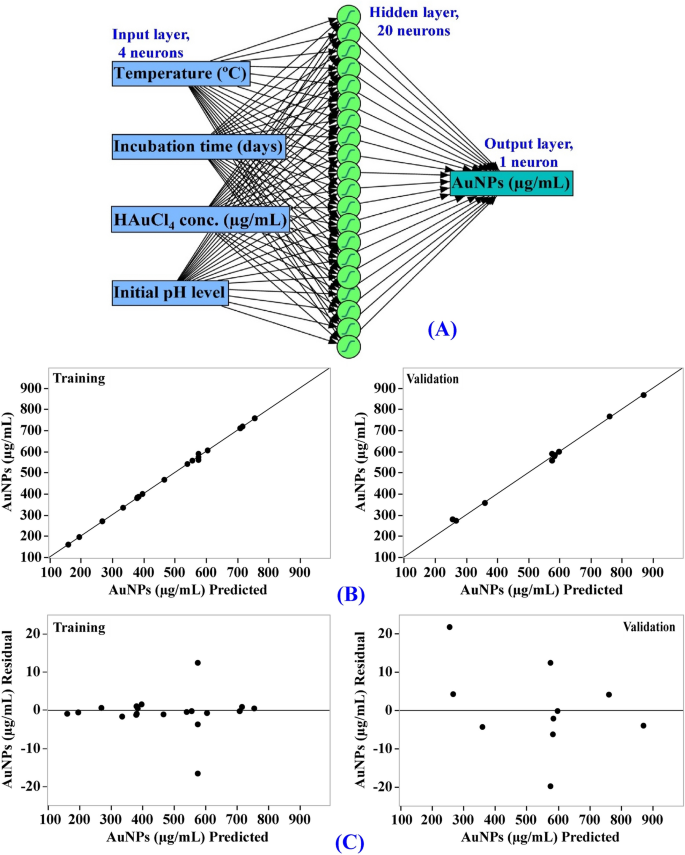
Enter neuron community topology was used to construct an ANN structure for optimizing AuNPs biosynthesis utilizing the cell-free supernatant of Streptomyces flavolimosus biomass. Easy neural community structure has interconnected synthetic neurons in three layers together with output layer, hidden layers and enter layer. On this research, one enter layer comprised of the 4 unbiased components (temperature (°C), incubation interval (days), HAuCl4 focus (µg/mL) and preliminary pH degree) enters the factitious neural community. Enter nodes course of the info, analyze or categorize it, and cross it to the following layer. One output layer offers the ultimate end result of the factitious neural community’s knowledge processing (AuNPs biosynthesis, µg/mL) (Fig. 7A). The optimum ANN parameters have been adjusted to variety of TanH nodes within the first layer (20), a validation methodology (holdback proportion, 0.2), a studying fee of 0.1, variety of excursions (2000), and becoming choice was reworking covariates.
Analysis of ANN mannequin
The values predicted AuNPs biosynthesis by ANN corresponding to every experimental end result got in Desk 1. The anticipated values of AuNPs biosynthesis by ANN have been drawn towards the precise values (Fig. 7B). In each the coaching and validation phases, the factors are gathered nearer to the road displaying the optimum prediction, indicating that the mannequin is dependable. The scattering of residual knowledge above and beneath the regression line revealed a standard scattering of the residuals (Fig. 7C) which assist for the suitability of the ANN mannequin.
Comparability of prediction potential of ANN versus CCD
As illustrated in Desk 1, the AuNPs biosynthesis values predicted by ANN exhibit a extra affordable settlement with the experimental end result, and the residuals values have been decrease than these obtained by the CCD mannequin. The mannequin’s efficiency was in contrast utilizing the mannequin comparability dialog within the JMP Pro16. For comparability, a number of error capabilities, in addition to the R2 used to judge the prediction potential of the CCD and ANN. Probably the most incessantly employed capabilities for the comparability have been R2, common absolute error (AAE) and root common squared error (RASE) for every regression mannequin that are proven in Desk 4. The prediction potential of the CCD and ANN was in contrast, the upper worth of R2 (0.9981) together with decrease worth for RASE (7.64) and decrease worth for AAE (4.60) (Desk 4) confirms ANN as the most effective mannequin with a better predictive capability for the optimum ranges of various physicochemical variables for AuNPs biosynthesis. This discovering could be defined by the aptitude of ANN to provide good efficiency, as coaching of the neurons was repeated a number of instances for varied physicochemical variables.
Within the laboratory, the CCD and ANN fashions for maximizing AuNPs biosynthesis utilizing Streptomyces flavolimosus cell-free supernatant have been validated utilizing the estimated predicted values of the 4 examined variables. With a purpose to validate each CCD and ANN fashions, the optimum estimated predicted values that maximize AuNPs biosynthesis utilizing Streptomyces flavolimosus cell-free supernatant have been calculated to be a focus of HAuCl4 that was 1000 μg/mL, an preliminary pH of 6, a temperature of 35 °C, and an incubation time of 4 days. Based mostly on CCD and ANN knowledge evaluation, the theoretical predicted AuNPs biosynthesis was 845.4 μg/mL and 870.36 μg/mL, respectively. The theoretical predicted values have been assessed experimentally. Beneath these circumstances, the utmost experimental worth of AuNPs biosynthesis utilizing Streptomyces flavolimosus cell-free supernatant was 866.29 μg/mL. The verification confirmed a excessive precision diploma of each fashions. Nevertheless, the theoretical predicted worth of AuNPs biosynthesis by ANN (870.36 μg/mL) was significantly nearer to the experimental worth (866.29 μg/mL) than that predicted by CCD (845.4 μg/mL), which signifies that ANN has stronger prediction potential compared to the CCD.
Desirability operate (DF)
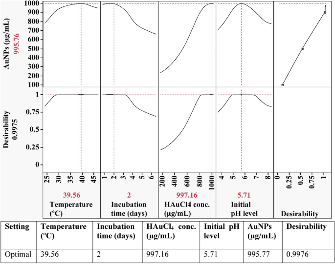
The desirability operate, proven in Fig. 8, was carried out with a purpose to discover the optimum predicted circumstances for biosynthesis of gold nanoparticles that will yield the very best worth.
The software program Design Knowledgeable’s desirability operate is about for any issue from 0 (undesirable) to 1 (fascinating)69. Usually, the worth of the desirability operate is estimated theoretically earlier than experimental verification of the optimization course of. The utmost worth of gold nanoparticles biosynthesis (995.77 µg/mL) utilizing the cell-free supernatant of Streptomyces flavolimosus was discovered beneath the optimum circumstances: HAuCl4 focus (997.16 µg/mL), preliminary pH degree 5.71, temperature 39.56 °C and incubation time 2 days. Beneath these circumstances, the utmost worth of gold nanoparticles biosynthesis utilizing the cell-free supernatant of Streptomyces flavolimosus (900.53 µg/mL) was confirmed, and the outcomes have been in comparison with the worth that had been predicted by the polynomial mannequin (995.77 µg/mL). The validation indicated a excessive degree of mannequin accuracy, demonstrating the mannequin’s validity on the issue ranges utilized.
Analysis of cytotoxicity and antitumor exercise in vitro
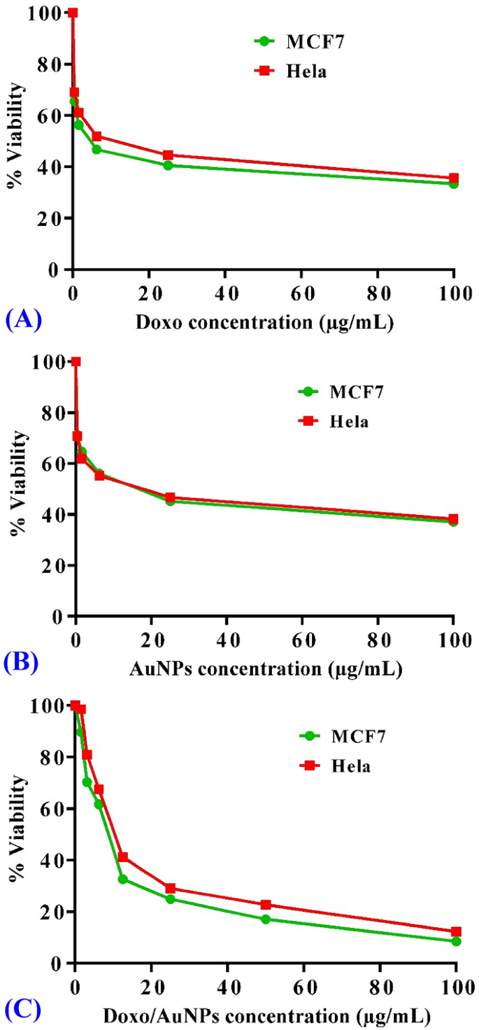
The anticancer efficacy (in vitro) of AuNPs synthesized by Streptomyces flavolimosus was evaluated in comparison with Doxorubicin (Doxo) utilizing MTT assay towards two human most cancers cell strains: cervical epithelioid carcinoma (Hela) and breast most cancers (MCF-7). Doxo considerably inhibited cell viability (Fig. 9A) in MCF-7 and Hela cells, with IC50 worth of 4.84 ± 0.28 μg/mL and 9.41 ± 0.31 μg/mL in MCF-7 and Hela cells, respectively. The obtained outcomes demonstrated that AuNPs exhibited a cytotoxic potential on the examined cell strains (Fig. 9B). As well as, remedies based mostly on AuNPs can destroy most cancers cells with an IC50 worth of 13.4 ± 0.44 μg/mL and 13.8 ± 0.45 μg/mL in MCF-7 and Hela cells, respectively. Furthermore, Doxo and AuNPs mixture considerably inhibited cell viability in MCF-7 and Hela cells, with IC50 worth of 8.32 ± 0.6 μg/mL and 11.72 ± 0.9 μg/mL in MCF-7 and Hela cells, respectively. Accordingly, addition of Doxo to AuNPs decreased considerably IC50 of AuNPs from 13.4 ± 0.44 μg/mL and 13.8 ± 0.45 μg/mL to eight.32 ± 0.6 μg/mL and 11.72 ± 0.9 μg/mL in MCF-7 and Hela cells; respectively. Thus, the mixture of Doxo and AuNPs elevated the anticancer potential of AuNPs supporting the additive impression of the anticancer potential when AuNPs and Doxo have been utilized in mixture. The rational of utilizing Doxo and AuNPs together is that the dose of every agent could be decreased to provide the identical required impact in addition to minimizing the unintended effects of Doxo. These outcomes counsel that AuNPs have the potential for use as an anticancer agent. Virmani et al.81 investigates the anticancer exercise of biogenic and chemically produced AuNPs in relation to malignant and regular cell strains. They noticed that the biologically produced AuNPs enormously suppressed the proliferation of malignant cells in a dose-dependent method with an IC50 worth of roughly 200 µg/mL. However, it was demonstrated that AuNPs will not be poisonous to regular cells. Myco-synthesized AuNPs made with the aqueous extract of the endophytic fungus Cladosporium sp. demonstrated anti-cancer efficacy towards MCF-7 by the induction of the apoptotic pathway82. IC50 worth was decided to 38.23 µg/mL. The research by Datkhile et al.83 evaluates the anticancer actions of AuNPs that have been biologically produced by using the extract of Nothapodytes foetida. The findings of the cytotoxicity take a look at demonstrated that the AuNPs had vital cytotoxic exercise towards most cancers cells; the IC50 worth have been 5.28, 7.2, and9.67 μg/mL for HCT-15, HeLa, and MCF-7 tumor cells, respectively.
Cytotoxicity of AuNPs in breast most cancers (MCF-7) and cervical epithelioid carcinoma (Hela) cell line. Focus-response plots of remedy with Doxo (constructive management) in MCF-7 and Hela cell strains (A), concentration-response plots of remedy with AuNPs in MCF-7 and Hela cell strains (B), and concentration-response plots of remedy with Doxo/AuNPs mixture in MCF-7 and Hela cell strains (C). The experiment was repeated three unbiased instances. Information are expressed as means ± SEM.
The anti-cancer results of AuNPs produced by Kaempferol have been examined on the MCF-7 most cancers cell line. The outcomes confirmed a dose- and time-dependent discount within the viability of MCF-7 breast most cancers cells84. In response to Kalaivani et al.85, AuNPs biosynthesized utilizing chitosan exhibited cytotoxic exercise towards MCF-7 cell strains with an IC50 worth of 250 μg/mL. Clarance et al.86 investigates the anticancer potentials of the AuNPs that have been produced by a inexperienced synthesis strategy with an endophytic pressure of Fusarium solani. They discovered that AuNPs displayed a dose-dependent cytotoxic impact on MCF-7 and HeLa cells. IC50 worth was decided to be 0.8 ± 0.5 μg/mL and 1.3 ± 0.5 μg/mL on MCF-7 and HeLa cell strains, respectively. Asl et al.87 examine the antitumor exercise of biogenically produced AuNPs on cisplatin-resistant A2780CP ovarian carcinoma cells. In a time- and dose-dependent method, AuNPs suppressed the proliferation of cisplatin-resistant ovarian carcinoma cells. Additionally, Abdulateef et al.88 examine the cytotoxic impact of the AuNPs synthesized utilizing bovine serum albumin and pulsed laser ablation method in simulated physique fluid. They discovered that the remedy had no impact on regular fibroblast cells. Nevertheless, a dose-dependent inhibition impact is noticed on the expansion of cervix most cancers cells (HeLa).
A number of research have demonstrated that the cytotoxicity of AuNPs is attributable to the induction of oxidative stress. HeLa cervical most cancers cells displayed enhanced reactive oxygen species (ROS) technology and oxidative stress when uncovered to 1.4 nm AuNPs, ensuing within the oxidation of proteins and lipids, a extreme impairment of the mitochondrial operate, and in the end cell loss of life89. Dickson et al.90 examine the cytotoxic impact of AuNPs that have been produced through inexperienced sources on HeLa and melanoma tumor cells. The outcomes confirmed that the cytotoxicity was dose-dependent and the impression of AuNPs on cell viability have been attributable to the induction of apoptosis.
The impact of AuNPs and doxorubicin collectively on cell viability on MCF-7 and Hela cells was checked. The outcomes confirmed that Doxo and AuNPs mixture considerably inhibited cell viability in MCF-7 and Hela cells, with IC50 worth of 17.71 ± 1.2 μg/mL and 23.11 ± 1.7 μg/mL in MCF-7 and Hela cells, respectively. Thus, this may be understood as an additive impression of the anticancer potential when AuNPs and Doxo have been utilized in mixture. The concentration-response of remedy with Doxo/AuNPs mixture in MCF-7 and Hela cell strains is proven within the Fig. 9C.
Aboyewa et al.91 examined the cytotoxicity of the co-treatment in MDA-321, KMST-6, HT-29, HeLa and Caski cell strains. They investigated the mechanistic results of cytotoxicity induced by the co-treatment of Caco-2 cells with biogenic AuNPs and DOX. Aboyewa et al.91 counsel that these biogenic AuNPs can function drug sensitizers when utilized in mixture with chemotherapeutic medication and improve their efficacy at a really low dosage. They reported that co-treatment of Caco-2 cells with DOX and the biogenic AuNPs enhances the medication’ results and efficacy by means of a synergistic anti-cancer mechanism. Moreover, they reported that the biogenic AuNPs have been successfully taken up by the most cancers cells, which, in flip, could have enhanced the sensitivity of Caco-2 cells to DOX. Furthermore, the mixture of DOX and the biogenic AuNPs inhibited the long-term survival of Caco-2 cells, decreased the manufacturing of reactive oxygen species (ROS), induced apoptosis, elevated mitochondrial depolarization and precipitated a speedy depletion of ATP ranges.
Evaluation of antitumor exercise of AuNPs in vivo in EAC mannequin
Following the environment friendly induction of Ehrlich stable tumors and as soon as the tumor quantity reached 100–200 mm3, the mice have been randomly divided into 4 teams to obtain remedy for 21 days. Mice bearing stable tumors have been handled with AuNPs (5 mg/kg/day) as single remedy or together with Doxo.
Affect of AuNPs on tumor quantity in mice carrying EAC
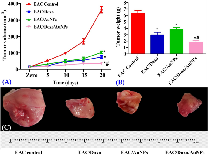
Analysis of tumor quantity each 5 days throughout the experiment demonstrated a major decline in tumor progress. On the finish of the experiment, the common tumor quantity within the EAC management mice had elevated from 145.6 to 3623 mm3. After 21 days of remedy, compared to EAC management mice, the mice that obtained Doxo and AuNPs remedy confirmed tumor progress inhibition fee of 79.2% and 71.6%; respectively; indicating a major antitumor exercise of AuNPs in EAC bearing mice. Whereas remedy with a mixture of Doxo and AuNPs resulted in a 90.5% tumor progress inhibition fee in comparison with EAC management group after 21 days of remedy suggesting that remedy with each Doxo and AuNPs on the similar time was simpler at decreasing tumor quantity than remedy with both drug alone (Fig. 10A).
Results of AuNPs alone or together with Doxo on tumor quantity (nm3) (A) and tumor weight (g) (B) of EAC bearing mice. All tumor photographs (C) have been taken on the similar magnification energy, zooming and distance from digital camera. Information are expressed as means ± SEM (n = 6 for EAC management group and n = 7 for EAC/Doxo, EAC/AuNPs, and EAC/Doxo/AuNPs teams). Statistical comparisons have been made utilizing a technique of study of variance (ANOVA) adopted by Tukey–Kramer put up hoc take a look at for a number of comparisons. *Signifies vital distinction vs. EAC management at P < 0.05; #represents vital distinction vs. EAC/AuNPs at P < 0.05.
Affect of AuNPs on tumor weight in mice carrying EAC
Evaluation of Ehrlich carcinoma weight throughout the experiment demonstrated a major decline in tumor weight. After 21 days of remedy, compared to EAC management mice, the mice that obtained Doxo and AuNPs remedy confirmed that the tumor weight decreased by 53.33% and 39.29%; respectively; indicating a major antitumor exercise of AuNPs in EAC bearing mice. Compared, remedy with a mixture of AuNPs and doxorubicin led to a lower in tumor weight that was 71.39% in comparison with that of the EAC management group (Fig. 10B,C). This vital tumor suppression within the group that was handled with the mixture of AuNPs and doxorubicin demonstrates that the anticancer efficacy of Doxo is considerably elevated when mixed with AuNPs.
In such areas of research, the appliance of AuNPs is attracting curiosity owing to their distinctive traits in comparison with the very poisonous NPs. AuNPs have wonderful conductivity, size-dependent traits, optical traits, low toxicity, simplicity, and useful variety. AuNPs are thought of to be comparatively biologically non-reactive and subsequently appropriate for in vivo purposes. The sturdy optical traits of sunshine absorbance of AuNPs as a consequence of localized floor plasmon resonance, enhanced permeability and retention in tumor tissue, their interplay with radiation to supply secondary electrons, in addition to their capability to be conjugated with medication or different medicines are additionally advantageous88,92,93. As a result of floor plasmon resonance attribute, laser stimulation of AuNPs induces hyperthermia and thermal harm of the tumor tissue whereas inflicting minimal hurt to regular tissue93. Myco-synthesized AuNPs made with the aqueous extract of the endophytic fungus Cladosporium sp. demonstrated sturdy cytotoxic efficacy towards the development of tumors in mice fashions carrying the cancerous tissue. Myco-synthesized AuNPs considerably decreased the physique weight, ascites quantity, and elevated the lifespan of EAC bearing mice82.
Affect of AuNPs on histopathological findings in EAC bearing mice
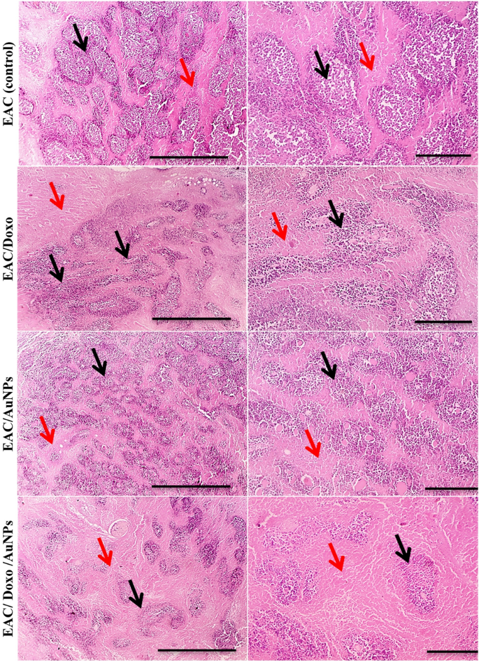
Microscopic photos of Hematoxylin and Eosin-stained pattern from EAC management mice displaying quite a few viable foci consisting of enormous, spherical and polygonal deeply stained tumor cells (black arrows) interspaced by small eosinophilic necrotic zones (white arrows) in subcutaneous and muscular tissues (Fig. 11). Variety of viable foci (black arrows) and variety of viable tumor cells are reasonably decreased related to reasonably elevated measurement of necrotic areas in group handled with Doxo. Variety of viable foci (black arrows), variety of viable tumor cells are barely decreased with conversely elevated measurement of necrotic areas (white arrows) in group handled with AuNPs. Variety of viable foci (black arrows) and variety of viable tumor cells are markedly decreased related to markedly elevated measurement of necrotic areas in group handled with a mixture of Doxo and AuNPs.
Microscopic photos of H&E-stained sections from tumor plenty displaying decrease viable foci consisting of enormous, spherical and polygonal deeply stained tumor cells (black arrows) interspaced by elevated eosinophilic necrotic zones (purple arrows) in handled teams with Doxo, AuNPs and mixture of Doxo and AuNPs in comparison with untreated EAC. A marked discount in tumor progress was noticed in group handled with mixture of Doxo and AuNPs. Magnifications × 40: bar 20 and × 100: bar 100.
As a result of AuNPs are a very good candidate to be used in biomedical purposes for photothermal remedy, biosensors, most cancers remedy, gene supply, drug supply, and imaging strategies, it’s important to judge their habits in an animal mannequin. A number of research point out that AuNPs accumulate in sure organs of rats94. A number of the components that affect the toxicity of AuNPs embody bodily dimensions similar to their measurement, dose, and form in addition to the size of publicity, their metabolism, floor chemistry, floor cost, composition, cell sort and immune response to the AuNPs95,96. The nanoparticles administration methodology may have an effect on toxicity, the injection of the nanoparticles by means of tail vein was much less poisonous than intraperitoneal and oral administration96. Moreover, the manufacturing methodology (chemical, bodily, and organic strategies) of the nanoparticles could possibly be a major issue within the toxicity results of the nanoparticles94.
Yahyaei et al.94 consider the affect of the biologically ready 50–70 nm AuNPs in vitro and in vivo in intraperitoneally injected rats. MTT assay outcomes revealed that AuNPs had in some way poisonous results which rely upon their doses. The in vitro and in vivo behaviors of the produced AuNPs have been completely different and AuNPs even in excessive focus induced low modifications within the rat organs. This can be because of the quick publicity and the usage of the biologically produced AuNPs. Shukla et al.97 reported that low concentrations of gold nanoparticles don’t end in a noticeable discount in physique weight or substantial toxicity. Crimson blood cells, hematocrit, and physique weight all decreased in excessive gold nanoparticle concentrations. Yah98 reported that AuNPs with a diameter of roughly 18 nm can enter cells with out inflicting cell harm or toxicity. Consequently, the small measurement of the AuNPs performs a major position and facilitates their incorporation into organic methods for subsequent probing and modification. Some exhibit toxicity because of the presence of surface-coated ligands, whereas others, as a consequence of their massive surface-to-volume ratio, function platforms for rising floor particle exercise. Moreover, it has been demonstrated that the affect of AuNP toxicity varies based mostly on particle form. Among the many shapes, it has been reported that rod-shaped AuNPs are extra poisonous than their spherical counterparts. There’s some data relating to the size-dependent elimination of chemically produced AuNPs from animal blood. As an example, it was reported that AuNPs with sizes of roughly 18 nm have been faraway from the bloodstream and gathered within the spleen and liver99. In distinction to the bigger AuNPs (50–100 nm), the smaller ones (5–15 nm) can simply be distributed all through the rat organs100. Yahyaei et al.94 demonstrated that short-term publicity to AuNPs had little impact on the liver and kidneys. However, short-term publicity to chemically produced AuNPs (50 nm) had negligible results on liver enzymes and no results in any respect on kidney enzymes101.
To keep away from accumulation and subsequent toxicity, nanoparticles (NPs) have to be metabolized or eradicated from the physique. The liver and the kidneys are the 2 main pathways by means of which NPs are eradicated from the physique. The mononuclear phagocyte system or tissue-resident phagocytes catch NPs within the hepatic pathway, resulting in their accumulation within the liver and spleen. The NPs are then subjected to biliary clearance and excretion through faeces and renal filtration route and eradicated by means of the urine102. On the mobile degree, it was demonstrated that AuNPs uptake occurred through receptor-mediated endocytosis, with most uptake occurring when nanoparticle sizes have been roughly 50 nm103. Hainfeld et al.104 demonstrated that small-sized AuNPs can cross the blood–mind barrier, and that this penetration could be managed by ion channel blockers. They demonstrated that there have been no opposed results on the brains of the animals and recommended utilizing AuNPs for drug supply functions.
