TPA NPs preparation and characterization
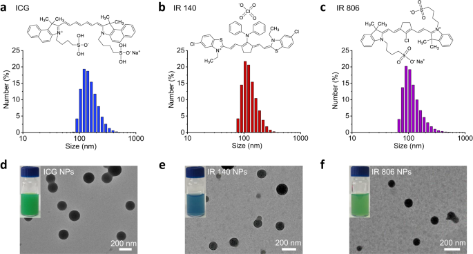
Supramolecular self-assembly has been extensively used as a technique to assemble NPs with novel optical properties35,36. On this regard, amino acid derivatives are versatile constructing blocks as a result of their ease of synthesis and huge number of physicochemical options, and have thus attracted growing consideration for the design of those NPs37. On this examine, the histidine (His) spinoff Z-His-Obzl (ZHO) (Supplementary Fig. 1), was chosen because the template amino acid spinoff to induce NIR cyanine dye ICG co-assembly as a result of its amphiphilic and positively charged nature. After mixing ICG aqueous answer with the ZHO dimethyl sulfoxide (DMSO) answer, a right away turbid answer occurred. The stoichiometry of ICG and ZHO was chosen as 0.125 mM and a couple of.020 mM, respectively, to realize optimized encapsulation effectivity (EE, 96.9%) and loading effectivity (LE, 11.2%) of ICG molecules (Supplementary Tab. 1). Nevertheless, transmission electron microscopy (TEM) picture confirmed the turbid answer consisted of nanodroplets (NDs), termed as ICG NDs, that had been kinetically-trapped unstable aggregates38 (Supplementary Fig. 2), additional evidenced by the presence of crystalline precipitates after growing old for twenty-four h (Supplementary Fig. 3). To stabilize the assembled nanostructures, zinc ions (Zn2+) had been launched to coordinate with the nitrogen atom of the imidazole group of ZHO to acquire extra thermodynamically favorable ICG NPs, as evidenced by the Fourier remodel infrared spectroscopy (FTIR) spectra consequence (Supplementary Fig. 4). In comparison with the ZHO and ICG, the vibration of imidazole teams in ICG NPs shifted from 1693 cm−1 to 1718 cm−1, indicating the coordination of Zn2+ with imidazole39. As anticipated, the colloidal stability of the ensuing ICG NPs was enhanced after growing old for twenty-four h (Supplementary Fig. 3) and the focus of Zn2+ was decided to be 0.438 mM. As well as, there was little distinction between the ICG NDs and the ICG NPs by way of spectral options, indicating an identical association of the ICG molecules (Supplementary Fig. 5). The improved absorbance depth of the ICG NPs additional prompt that Zn2+ coordination improves structural robustness40 (Supplementary Fig. 5). The obtained ICG NPs possess common hydrated diameters of 159.8 ± 48.1 nm (Fig. 2a and Supplementary Tab. 2), as decided by dynamic mild scattering (DLS). To display the universality, different NIR cyanine dyes, together with IR 140 and IR 806 had been co-assembled by the identical methodology. The typical sizes of IR 140 NPs and IR 806 NPs had been discovered to be 132.2 ± 42.1 nm (Fig. 2b and Supplementary Tab. 2) and 123.5 ± 56.7 nm (Fig. 2c and Supplementary Tab. 2), respectively. TEM photos additional confirmed the spherical morphologies of ICG NPs, IR 140 NPs and IR 806 NPs (Fig. 2nd–f), with sizes that had been almost an identical to their DLS information.
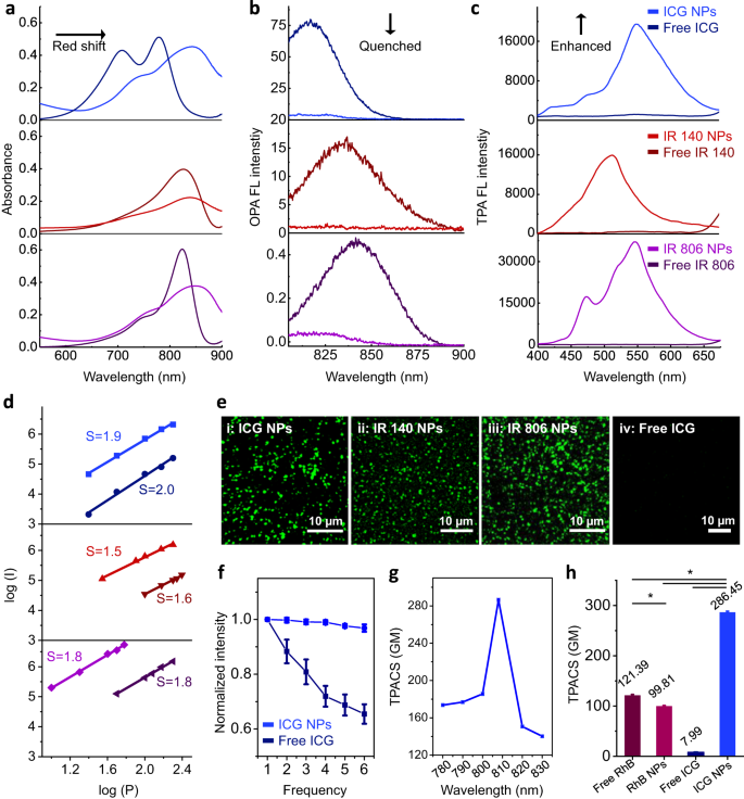
Subsequently, the group of the chromophores inside the nanoparticles was investigated. In contrast with their free state, NIR cyanine dye-based NPs confirmed a broadened and enormous red-shift within the absorption spectra (Fig. 3a), which could be attributed to the electron delocalization promoted by noncovalent interactions41. Of notice, such intermolecular interactions considerably decreased their one-photon absorption (OPA) fluorescence depth (810–900 nm), the place the calculated fluorescence quenching effectivity of ICG NPs, IR 140 NPs and IR 806 NPs was 93.8%, 93.4% and 92.2%, respectively (Fig. 3b). As one other fundamental pathway of photo-activated NIR cyanine dyes42, photothermal rest of NPs was inferior than the corresponding free dyes as effectively (Supplementary Fig. 6). Remarkably, in response to femtosecond (fs) Ti: Sapphire oscillator 808 nm laser irradiation, NIR cyanine dye-based NPs exhibited drastically enhanced TPA fluorescence emission (400–650 nm) (Fig. 3c). Comparable habits was noticed for all three investigated NIR cyanine molecules, however not for different dyes together with porphyrins (protoporphyrin IX (PpIX), tetraphenylporphyrin tetrasulfonic acid (TPPS)) and phthalocyanines (nickel(II) phthalocyanine-tetrasulfonic acid tetrasodium salt (NiTSPc), naphthalocyanine (NaPc)) (Supplementary Fig. 7). All NPs had been ready with the identical methodology and their TPA fluorescence was quenched when evaluating with their free state. These comparative outcomes prompt that the improved TPA fluorescence could be a singular property of NIR cyanine dyes. To know the elevated fluorescence upon TPA, this course of was investigated in additional element. By altering the facility power, the TPA fluorescence spectra of free NIR cyanine dye-based NPs and the corresponding free dyes had been recorded (Supplementary Fig. 8). The fluorescence of free NIR cyanine dyes displayed quadratic emission depth as a perform of elevated incident energy power (Fig. 3d and supplementary Tab. 3), suggesting that the resonance construction of the cyanine dyes promoted the inherent push-pull electron switch for TPA. Importantly, as soon as NPs had been fashioned, the TPA fluorescence depth elevated not less than one order of magnitude compared with free NIR cyanine dyes (Supplementary Fig. 8 and Fig. 3d), implying that the aggregated state enhanced the TPA fluorescence emission. Furthermore, the TPA fluorescence emission was noticed by using a confocal laser scanning microscope (CLSM) geared up with a fs Ti: Sapphire oscillator laser on the wavelength of 808 nm and an emission channel of 495-540 nm, which lined the spectral emission starting from 400 to 650 nm of the NIR cyanine dye-based NPs (Fig. 3e: i-iii). To display the significance of the aggregated state within the commentary of the improved TPA and the broad applicability of this idea, we constructed a sequence of various NIR dye aggregates and in contrast them to the free state (Fig. 3e: iv). In addition to, we obtained the dye complexes with proteins and polypeptides: ICG/bovine serum albumin (BSA) advanced (Supplementary Figs. 9a and 10a), IR140/BSA NPs (Supplementary Figs. 9b and 10b) and ICG/poly(L-lysine) (PLL) NPs (Supplementary Figs. 9c and 10c). All assemblies confirmed enhanced TPA fluorescence, confirming that the aggregated state is an important function to facilitate enhanced TPA fluorescence emission of cyanine dyes.
a Absorption spectra, b OPA fluorescence (FL) spectra and c TPA FL spectra of free NIR cyanine dyes and their corresponding NPs. d Linear curve fitted by double exponential mannequin between the emission depth log (I) and excitation energy log (P), the place the “S” indicated the slope of fitted strains. e CLSM photos of (i) ICG NPs, scale bar is 10 µm; (ii) IR 140 NPs, scale bar is 10 µm; (iii) IR 806 NPs, scale bar is 10 µm and (iv) free ICG, scale bar is 10 µm. All figures are obtained within the emission vary of 495-540 nm. f The photo-stability take a look at of free ICG and ICG NPs as a perform of scanning frequency. The ability density of laser excitation was 1.5 W cm−2. Error bars denote the usual deviation (n = 3 impartial experiments). Information are offered as imply values +/− S.D. g TPACS of ICG NPs at completely different wavelengths utilizing RhB dissolved in methanol as reference. Error bars denote the usual deviation (n = 3 impartial experiments). Information are offered as imply values +/− S.D. h Comparability of TPACS of free RhB, RhB NPs, free ICG and ICG NPs. Error bars denote the usual deviation (n = 3 impartial experiments). Information are offered as imply values +/− S.D., and P values are calculated by one-way ANOVA *P < 0.05. The ability density of laser excitation for all TPACS measurements was 0.1 W cm−2. The focus of NIR cyanine dyes and RhB utilized in all figures was 25 µM.
Because of the constructive impact of nanostructures in retaining the steadiness of emitted fluorescence43,44, photo-bleaching injury was considerably alleviated. Taking ICG NPs for example, superior anti-photo-bleaching stability, moderately than fast degradation as noticed free of charge ICG, was noticed (Fig. 3f). Additionally, the introduction of Zn2+ elevated the TPA fluorescence stability (Supplementary Fig. 11). These outcomes had been primarily ascribed to structural safety related to the formation of steady nanoarchitectonics. To achieve a deeper perception into the extremely uncommon TPA optical properties of NIR cyanine dye-based nano-assemblies, comparative outcomes of ICG with the natural fluorescent dye RhB (Supplementary Fig. 12a), generally used as TPA fluorophore, had been obtained. Ready in the identical method because the ICG NPs, RhB NPs possessed a uniform spherical morphology with a mean diameter of 136.5 ± 51.3 nm (Supplementary Fig. 12b and c) and confirmed a broadened absorption peak with spectroscopy that was indicative of meeting formation (Supplementary Fig. 12d). Moreover, a quenching impact in OPA fluorescence (Supplementary Fig. 12e) was noticed. The TPA fluorescence spectra of RhB with variable excitation wavelength confirmed that the TPA fluorescence emission of RhB was positioned within the vary of round 520–680 nm when concurrently absorbing two photons and was impartial of the excitation wavelength used. This commentary indicated that somewhat bit power loss was inevitable within the means of power transition from excited state to floor state, thus resulting in the deviation of the emitted photon from the theoretical power with 2-fold frequency1 (Supplementary Fig. 12f). When topic to the excitation wavelength of 808 nm, it was discovered that the assembled RhB NPs confirmed somewhat red-shifted emission and barely decreased depth as compared with free RhB (Supplementary Fig. 12g and h). This may be contributed to the ACQ impact of RhB, completely different from the noticed phenomenon of enhanced TPA fluorescence in NIR cyanine dye. Additional, the TPACS (δ), as essentially the most generally used parameter with a unit of Goeppert-Mayer (GM, 1 GM = 10−50 cm4·s photon−1) for characterizing TPA chromophores45, was quantitatively measured. ICG NPs confirmed a wavelength-dependent habits and gave a most of 286.45 GM upon 808 nm laser excitation (Fig. 3g), which is 2.36-fold and a couple of.87-fold greater than free RhB and RhB NPs, respectively. Importantly, the TPACS of ICG NPs was 35.90-fold bigger than free ICG (Fig. 3h and supplementary Tab. S4), indicating that ICG NPs are extra helpful in TPA fluorescence imaging compared to free ICG.
Picture-oxidation enhanced emission mechanism of TPA NPs
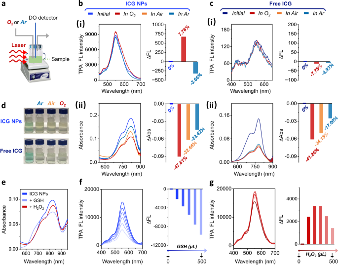
Additional research had been performed to uncover the underlying mechanism governing the improved TPA fluorescence emission. Intriguingly, the fluorescence emission of ICG NPs and free ICG confirmed completely different habits upon photo-oxidation (Fig. 4a). The fluorescence depth upon laser irradiation of ICG NPs was enhanced, ∆FL/FLpreliminary = 7.67%, in aqueous answer saturated with O2 (dissolved oxygen focus of 11.5 mg mL−1), whereas this transformation was reverse in aqueous answer saturated with Ar (dissolved oxygen focus of three.6 mg mL−1) with ∆FL/FLpreliminary = −3.66% (Fig. 4b-i). In distinction, solely photo-degradation was noticed when free ICG was irradiated beneath these completely different oxygen-containing circumstances (In O2-rich situation: ∆FL/FLpreliminary = −7.75%; In Ar-rich situation: ∆FL/FLpreliminary = −4.93%) (Fig. 4c-i). In absorption spectroscopy, a shade change of free ICG from inexperienced to yellow was noticed and the loss in absorbance was proportional to the dissolved oxygen focus: the ∆Abs/Abspreliminary of free ICG in Ar-rich situation is −17.00% whereas its ∆Abs/Abspreliminary in air situation and O2-rich situation is −34.15% and −41.26%, respectively (Fig. 4c-ii and d), confirming the photo-degradation phenomenon. An analogous tendency in absorption spectra was noticed for ICG NPs: the ∆Abs/Abspreliminary of ICG NPs in Ar-rich situation is −23.42% whereas its ∆Abs/Abspreliminary in air situation and O2-rich situation is −32.66% and −47.81%, respectively (Fig. 4b-ii and d), which thus differed from their fluorescence depth enhancement. The inconsistency between fluorescence and absorbance motivated us to conjecture that the improved fluorescence emission is mediated by short-lived unstable intermediates. Alternatively, the TPA fluorescence could be simply managed by different means, together with addition of an O2 scavenger (glutathione, GSH) in addition to elevating oxygen ranges (addition of H2O2), that confirmed little affect on the assembled nanostructures (Fig. 4e). As displayed in Fig. 4f, the fluorescence emission of ICG NPs was diminished when GSH depleted dissolved oxygen or associated lively species, and their ∆FL/FLpreliminary was modified to −63.05% because the GSH quantity elevated as much as 500 μL. Not surprisingly, the fluorescence emission confirmed oxygen-dependent photo-oxidation enhancement to some extent upon addition of H2O2. When the amount of H2O2 was beneath 300 μL, the ∆FL/FLpreliminary of ICG NPs modified as 24.55% whereas when the amount of H2O2 exceeded 300 μL to 500 μL, i.e., when extreme photo-oxidation was induced, chromophores within the ICG NPs degraded as effectively as a result of their ∆FL/FLpreliminary decreased from 24.55% to 12.26% (Fig. 4g). These outcomes once more demonstrated the existence of oxygen-dependent unstable intermediates, which enhanced the TPA fluorescence.
a Schematic illustration of ICG NPs in numerous oxygen-dependent atmospheres. b (i) TPA FL spectra and the (ii) absorption spectra of ICG NPs by laser irradiation (1.5 W cm−2, 10 min) in numerous oxygen-dependent atmospheres. c (i) TPA FL spectra and the (ii) absorption spectra of free ICG by laser irradiation (1.5 W cm−2, 10 min) in numerous oxygen-dependent atmospheres. The proper in Fig. b (i) and c (i) indicated the ∆Fluorescence (∆FL) with the definition of FL−FLpreliminary, and the worth of ∆FL/FLpreliminary was famous. The proper in Fig. b (ii) and c (ii) indicated the ∆Abs with the definition of Abs−Abspreliminary, and the worth of Abs/Abspreliminary was famous. d Optical footage of ICG NPs and free ICG after laser irradiation (1.5 W cm−2, 10 min). e Absorption spectra of ICG NPs after including reductant (GSH) and oxidant H2O2. TPA fluorescence spectra of ICG NPs as a perform of f GSH and g H2O2. The focus of ICG utilized in all figures was 25 µM.
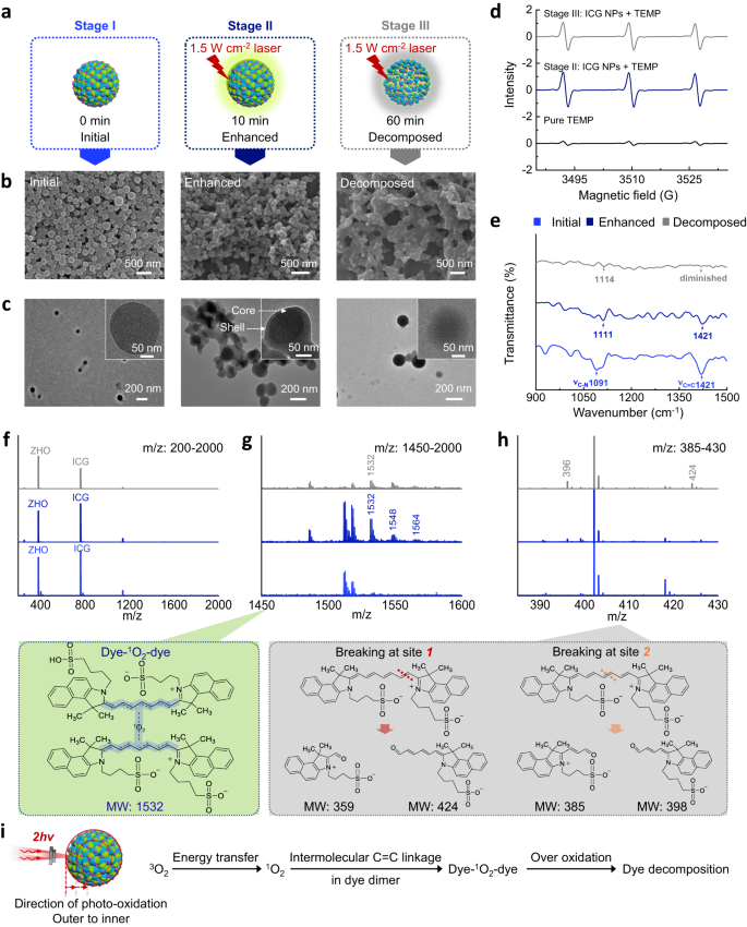
With a view to make clear the properties and construction of the intermediates, the fluorescence modifications of ICG NPs had been categorised into preliminary, enhanced and decomposed levels in keeping with the photo-oxidation course of. The preliminary stage (termed as stage I) refers back to the ICG NPs with out laser irradiation. The improved stage (termed as stage II) refers to when ICG NPs had been irradiated by laser (1.5 W cm−2, 10 min) to indicate the improved fluorescence. The decomposed stage (termed as stage III) means when no fluorescence of ICG NPs was detected after laser irradiation (1.5 W cm−2, 60 min) (Fig. 5a). When it comes to morphology characterised by scanning electron microscope (SEM) (Fig. 5b) and TEM (Fig. 5c), spherical nanostructures with uniform measurement distribution had been noticed in stage I. As soon as photo-oxidation occurred and accompanied with enhanced TPA fluorescence emission, NPs appeared extra adherent and crossed collectively. Moreover, in stage II, the constructions advanced from spherical to core-shell. In stage III (extreme photo-oxidation), crossing between NPs additional elevated and core-shell constructions disappeared. Based mostly on the noticed morphology modifications within the photo-oxidation course of, it may be inferred that degradation proceeded step by step from the surface to the within of ICG NPs, and unstable intermediates answerable for enhanced TPA fluorescence emission could also be generated as effectively. Clearly, oxygen is essential within the photo-oxidation enhanced fluorescence emission and determines the construction of intermediates of ICG NPs. Due to this fact, the practical type of oxygen was detected by electron paramagnetic resonance (EPR). The 1O2 detector probe 2,2,6,6-tetramethyl-piperidin (TEMP), was used to react with 1O2 to type a steady nitroxide radical product 2,2,6,6-tetramethylpiperidine-1-oxyl (TEMPO), which could be recorded by EPR46. As proven in Fig. 5d, in contrast with the technology of TEMPO sign within the pure TEMP group, the depth of TEMPO sign considerably elevated in stage II. This prompt that the presence of 1O2 in ICG NPs promoted the formation of TEMPO. Importantly, the TEMPO sign depth in stage II was greater than that within the stage III, implying that 1O2 certainly participated within the photo-oxidation mediated the enhancement of TPA fluorescence emission. Moreover, FTIR spectra (Fig. 5e) and electrospray ionization mass spectra (ESI-MS) (Fig. 5f–h) of ICG NPs at completely different levels had been recorded. The sign of the C = C stretching vibration of ICG molecules47 diminished from stage I to stage III, implying that C = C bonds had been attacked by photo-oxidation. The bands comparable to the C-N stretching vibration could be detected in three levels, suggesting that C-N bonds weren’t affected by photo-oxidation (Fig. 5e). In ESI-MS, a attribute m/z of 1532 peak that was not noticed within the stage I, appeared in stage II and was nearly extinct in stage III (Fig. 5f and g). The m/z peaks, resembling 398 and 424, which indicated fragment merchandise by 1O2-mediated photo-degradation of ICG molecules at completely different C = C bonds48, had been noticed (Fig. 5h). Therefore, the molecules comparable to m/z = 1532 are intermediates that govern the improved fluorescence emission. Based mostly on this evaluation, a doable construction of those intermediates is listed in Fig. 5g, the place two ICG molecules are linked by 1O2 and the linking websites are positioned on the C = C bond. This 1O2-containing intermediate contributed to photo-oxidation enhanced TPA fluorescence of ICG NPs, a singular attribute of ICG NPs, fairly completely different from the ICG dimer with out connection by oxygen49. Molecular dynamics (MD) simulation outcomes (Supplementary Fig. 13) supported our speculation: ZHO mediated self-assembly of ICG resulted in nearer distance between neighboring ICGs. The ICG molecules exhibited a minimal intermolecular distance of roughly 3.3 Å, which facilitated electron delocalization for TPA fluorescence emission50 and paved the way in which for 1O2 to hyperlink their C = C bonds. Upon laser irradiation, ICG molecules that had been excited to the T1 state switch their power to 3O2 by forming 1O2. Then, 1O2 intermolecularly conjugated the C = C bonds to type intermediates (dye-1O2-dye) inside ICG NPs, additional enlarging electron delocalization for TPA absorption and eventually resulting in enhanced fluorescence emission (Fig. 5i). This mechanism offers a believable rationalization for the noticed enhancement of TPA fluorescence emission.
a Schematic illustration of preliminary, enhanced (1.5 W cm−2, 10 min) and decomposed (1.5 W cm−2, 60 min) levels of ICG NPs. Morphological characterizations of ICG NPs at completely different levels: b SEM photos and c TEM photos. d EPR spectra of ICG NPs at completely different levels. TEMP was added as indicating agent throughout laser irradiation. e FTIR transmittance spectra of ICG NPs at completely different levels. f–h ESI-MS of the ICG NPs and doable chemical constructions at completely different levels. Fig. f, g and h denote the m/z vary 200-2000, 1450-1600 and 385-430, respectively. i Schematic illustration of photo-oxidation enhanced emission mechanism. The focus of ICG utilized in all figures was 25 µM.
Imaging utility of TPA NPs
Given the truth that stability of NPs is essential and has been a formidable problem in biomedical imaging51, the steadiness of ICG NPs was investigated. In absorption spectroscopy (Supplementary Fig. 14), upon storage, the absorption peak of ICG NPs confirmed little alteration throughout 15 days of growing old (normalized 16.44% lower in depth). Against this, free ICG molecules underwent fast degradation (normalized 54.51% lower in depth). Moreover, a red-shift appeared on the 15th day, suggesting the free ICG was unstable and fashioned massive aggregates52. These comparative outcomes demonstrated that the a number of noncovalent interactions endowed the ICG NPs with colloidal stability. As well as, by creating 10-fold (v/v) dilutions of their suspensions with 10% fetal bovine serum (FBS) and incubating the suspensions at 37 °C for twenty-four h, the morphology of the ICG NPs confirmed no discernible change as an alternative of somewhat measurement improve assigned to serum protein adsorption (Supplementary Fig. 15 and supplementary Tab. 5), suggesting glorious physiological stability. Contemplating the acidic microenvironment of tumors, stability take a look at in mimicking tumoral acid-microenvironment was performed as effectively. The absorption spectra of ICG NPs and RhB NPs had been recorded in numerous pH circumstances to observe their stability (Supplementary Fig. 16). Other than a slight absorption lower, no band shift was noticed in ICG NPs when lowering pH worth from 7 to five. However a substantial turbidity lower and a blue-shift had been noticed in RhB NPs, indicating their disassembly53. The disassembly propensity of RhB NPs could be attributed to pH-susceptibility of RhB-based dyes54. Against this, the much less susceptibility of ICG NPs to pH variation highlighted their benefits in tumor imaging utility.
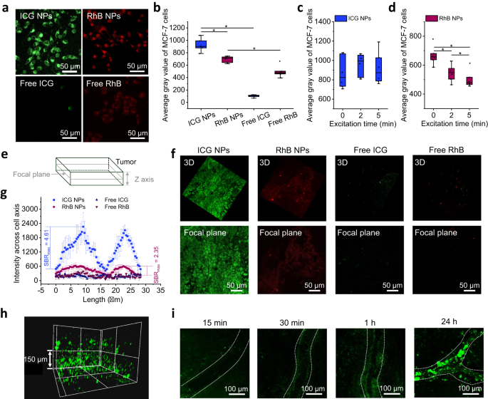
Subsequent, the in vitro and in vivo utility potentials of ICG NPs had been assessed by evaluating to free ICG, RhB NPs and free RhB. An ordinary 3-(4,5-dimethylthiazolyl-2)-2,5-diphenyltetrazolium bromide (MTT) cell survival assay relating to cytotoxicity of 4 completely different formulations was performed (Supplementary Fig. 17). Cell viability was not affected when incubating the cells with NPs on the focus vary from 0 µM to 200 µM. Furthermore, no vital distinction of cell viability was noticed between NPs group and their free state group. These outcomes indicated their considerable biocompatibility, paving the way in which for his or her organic utility. After incubating cells with free dyes and NPs for twenty-four h, CLSM photos (Fig. 6a and b) demonstrated that the dyes had been most definitely endocytosed within the cytoplasm, and the NPs (ICG NPs and RhB NPs) confirmed the a lot greater fluorescence depth than free dyes (free ICG and free RhB), suggesting that nanostructures elevated the endocytosis effectivity of dyes. By comparability, the fluorescence depth of RhB NPs within the cytoplasm was inferior to ICG NPs, which was attributed to their decrease δ worth and diminished stability in acidic circumstances. Moreover, the photo-bleaching take a look at relating to ICG NPs and RhB NPs was performed as effectively. Human breast most cancers MCF-7 cells had been repeatedly excited and their common grey was recorded at 0 min, 2 min and 5 min to point the fluorescence depth. Throughout steady photo-bleaching, ICG NPs within the mobile cytoplasm maintained sturdy photo-luminescence even after being irradiated as much as 5 min (Fig. 6c), which could be primarily attributed to their photo-oxidation enhanced fluorescence emission and physiological stability. In distinction, RhB NPs confirmed distinct behaviors as they had been step by step bleached over excitation time (Fig. 6d). Because the in vitro experiments confirmed sturdy TPA fluorescence emission and superior anti-photo-bleaching capbility, ICG NPs served as glorious candidates for the imaging implementation in vivo.
a Consultant CLSM photos and b calculated common grey values of MCF-7 cells that incubated with 4 sorts of formulations together with ICG NPs, free ICG, RhB NPs and free RhB (dye focus: 25 µM) for twenty-four h. Error bars denote the deviation (n = 6 biologically impartial cells). Information are offered as imply values +/− S.D., and P values are calculated by one-way ANOVA *P < 0.05. Calculated common grey worth of MCF-7 cells in c ICG NPs and d RhB NPs over excitation time (0, 2, 5 min). The ability density of laser excitation was 1.5 W cm−2. Error bars denote the deviation (n = 6 biologically impartial cells). Information are offered as imply values +/− S.D., and P values are calculated by one-way ANOVA *P < 0.05. e Schematic illustration of tumor tissue imaging in vivo. f Consultant three-dimensional reconstruction photos of tumor part alongside Z axis (higher) and focal airplane photos (decrease), the place the Z depth is 20 µm. Mice bearing MCF-7 tumor had been intravenously injected with ICG NPs, free ICG, RhB NPs and free RhB (dye dosage: 250 µM, 200 µL) for twenty-four h. The size bar is 50 µm. g Fluorescence depth cross cell axis of MCF-7 cells obtained from Fig. f. Error bars denote the deviation (n = 20 biologically impartial cells). Information are offered as imply values +/− S.D., and P values are calculated by one-way ANOVA *P < 0.05. h Three-dimensional reconstruction of tumor tissue after intravenous injection of ICG NPs for twenty-four h. The size bar is 150 µm. i Consultant CLSM photos of real-time readout means of ICG NPs accumulation in blood vessels inside tumor websites at completely different time intervals (15 min, 30 min, 1 h and 24 h). The white line signifies the blood vessel. The size bar is 100 µm.
The in vivo imaging was carried out in tumor-bearing mice. When the tumor quantity reached 150 mm3, NPs (ICG NPs, RhB NPs) and their free state formulations (free ICG and free RhB) with the identical dye dosage (250 µM, 200 µL) had been intravenously injected into mice. At 24 h post-injection, the mouse was anesthetized and the tumor website was imaged. From the focal airplane, decrease and higher sections had been scanned alongside the Z axis (Fig. 6e). As proven in Fig. 6f, the fluorescence sign obtained on the mobile stage could be obtained, the place the higher panel confirmed the three-dimensional reconstruction photos of tumor part with Z depth of 20 µm, and the decrease airplane confirmed the focal airplane photos. Cell entities could be recognized in teams of ICG NPs and RhB NPs, however not in free ICG and free RhB teams, suggesting that NPs amassed in tumor websites whereas free dyes could also be rapidly cleared from mice physique55. Importantly, the imaging of ICG NPs inside tumor cells was clearer than that of RhB NPs, verifying the extraordinary fluorescence emission in vivo of ICG NPs. Additionally, the TPA fluorescence imaging of ICG NPs has been demonstrated in different three sorts of mice xenograft tumor fashions together with mouse breast carcinoma cell line 4T1, mouse colorectal carcinoma cell line CT26 and human cervical most cancers cell line Hela (Supplementary Fig. 18), indicating pervasive utility of ICG NPs to different forms of tumors. Furthermore, the utmost signal-to-background ratio (SBRmax) cross cell axis, to characterize imaging distinction, was analyzed. The SBRmax of ICG NPs was calculated as 4.61, which is 1.96-fold greater than the counterpart worth of RhB NPs (SBRmax = 2.35) (Fig. 6g). In addition to, the three-dimensional scanning enabled the visualization of the distribution of ICG NPs in tumor tissue (Fig. 6h and supplementary Mov. 1), the place the size alongside the Z-axis reached round 300 µm. Intriguingly, a real-time readout of the presence of ICG NPs inside tumor vascular circulation was realized (Fig. 6i). Blood vessels in tumor tissue confirmed a comparatively darkish state about 15 min after intravenous injection. Over time, nevertheless, ICG NPs circulated to the tumor vascular websites and amassed on the tumor vascular partitions, which indicated an enhanced permeability and retention (EPR) impact. Additional, ex vivo imaging of tumor biopsies (Supplementary Fig. 19), demonstrated that ICG NPs had been endocytosed by cells with out damaging their morphology, predicting that ICG NPs could also be secure. Lastly, OPA fluorescence imaging was performed to match the property of TPA fluorescence. Data relating to the distribution of NPs in numerous intact tissues at 24 h postinjection could be collected, which additionally confirmed that the ICG NPs possessed EPR impact inside tumor (Supplementary Fig. 20). Extra detailed data, such because the mobile distribution inside tumor tissue, can’t be supplied by OPA fluorescence imaging approach. The comparisons emphasised the benefits of ICG NPs-based TPA fluorescence imaging: (i) The chromophores solely on the focus level could be excited in TPA fluorescence imaging, which is the inherent nature of TPA imaging approach and assured excessive imaging decision; (ii) The excessive distinction imaging on the mobile stage in vivo could be obtained owing to their excessive TPA cross-section, glorious stability, appreciable anti-photo-bleaching functionality and excellent SBR of ICG NPs.
